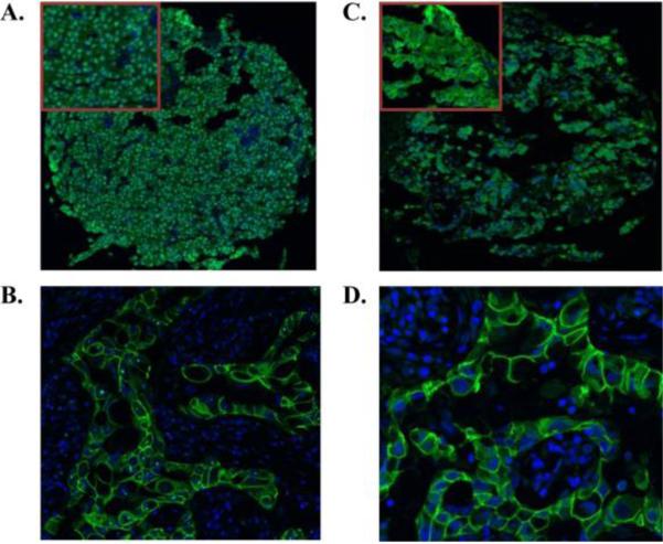
Figure 1.
Representative IHC of EGFR and ErbB2 receptors in prolactinomas on tissue array (20× magnification). Top corner box shows magnification of selected area. EGFR staining was nuclear and ErbB2 staining membranous/cytoplasmic. Positive control tissue is breast cancer A. EGFR staining of prolactinoma B. EGFR staining of breast cancer (63× magnification). C. ErbB2 staining of prolactinoma D. ErbB2 staining of breast cancer (63× magnification).

