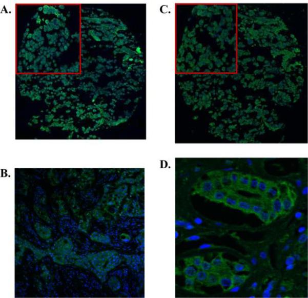
Figure 2.
Representative IHC of ErbB3 and ErbB4 receptors in prolactinomas on tissue array (20× magnification). Top corner box shows magnification of selected area. Positive control tissue is breast cancer for ErbB3 and kidney tubules for ErbB4. A. ErbB3 staining of prolactinoma B. ErbB3 staining of breast cancer (20× magnification) C. ErbB4 staining of prolactinoma D. ErbB4 staining of kidney tubules (63× magnification).

