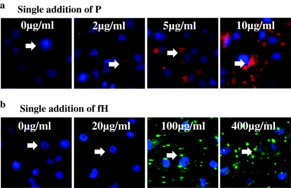
Figure 3.

Binding of properdin (P) and factor H (fH) on proximal tubular epithelial cells (PTECs). PTECs were incubated with variable concentrations of P (0–10 μg/mL) or fH (0–400 μg/mL) for 3 h. Dose-dependent depositions of P (a) or fH (b) were observed by immunofluorescence (IF). Depositions of P and fH were observed at a concentration (P, ≥2 μg/mL; fH, ≥20 μg/mL) lower than the physiological serum concentration (P, 4–6 μg/mL; fH, 300–500 μg/mL). Original magnification: ×100. Arrow: depositions of P or fH.
