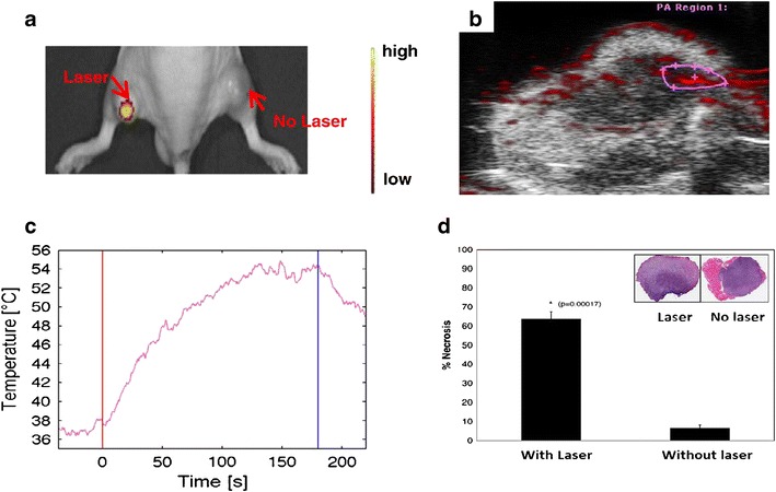
Fig. 5.

In vivo fluorescence intensity studies. a Representative in vivo fluorescence optical imaging of DOX release following the intratumoral injection of DOX@PEG-HAuNS at t = 24 h. b Overlaid photoacoustic and B-mode images of DOX release in vivo following intratumoral injection of DOX@PEG-HAuNS (1.32 × 10¹² particles/mL) and treatment with a 6-W surface laser. c Conversion of photoacoustic signal to temperature. The first vertical line indicates the start of laser treatment, and the second vertical line indicates the end of laser treatment. d Histological analysis. The percentage of tumor necrosis in laser-treated mice (64%) was significantly higher than that in untreated mice (7%). Inset representative hematoxylin and eosin-stained slides of 4T1 tumors injected intratumorally with DOX@PEG-HAuNS with and without NIR surface laser treatment (0.15 W/mm2 for 1 min). Reproduced with permission from ref. (41). Copyright 2013, Elsevier
