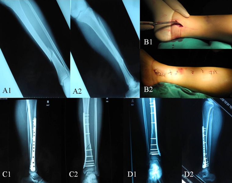
Fig. 1.
a1-a2 Plain radiographic anteroposterior and lateral views of a distal tibial shaft fracture. b1-b2 Minimally invasive surgery incision. c1-c2 Plain radiographic anteroposterior and lateral views two days after operation. d1-d2 Plain radiographic anteroposterior and lateral views 12 months after operation

