
Figure 6.
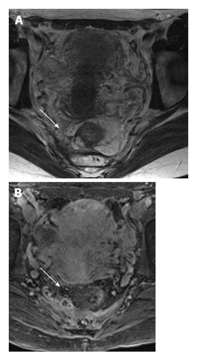
A 45-year-old female with fistula extending above the levator ani muscles (supralevator) and forming an abscess (white arrows). The signal intensity of the fat and fluid are the same on T2 without fat saturation, and the abscess can easily be missed on T2 weighted images (A). However, the marked inflammation on 3D T1 post-contrast clearly demonstrates the abscess (B).