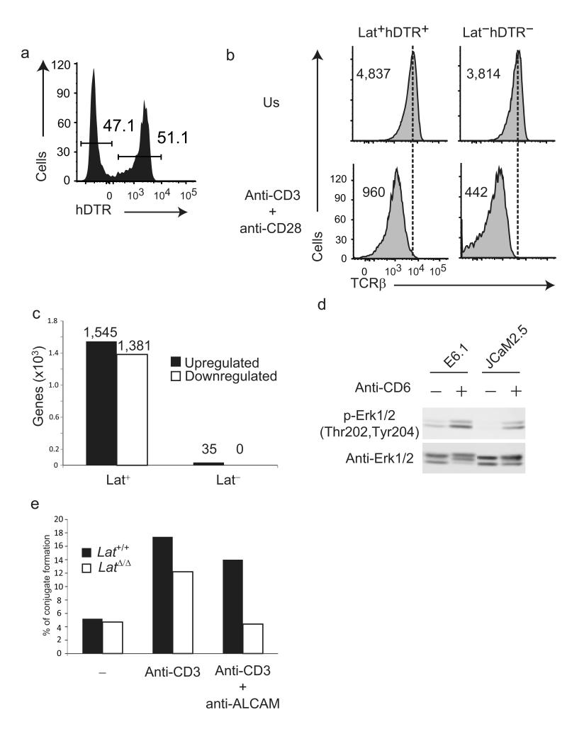
Figure 7.
Limited transcriptional changes occur when the TCR is engaged in the absence of Lat. (a) Expression of hDTR at the surface of CD4+ T cells of 4 week-old mice of maT-Cre x Latfl-dtr genotype. Percentages of Lat−hDTR− and Lat+hDTR+ CD4+ T cells are shown. (b) Expression of TCRβ on sorted Lat+hDTR+ and Lat−hDTR− CD4+ T cells that were stimulated for 4 h with anti-CD3 and anti-CD28 (anti-CD3 + anti-CD28) or left untreated (NS). Numbers indicate the geometric mean fluorescence. (c) Numbers of genes that were significantly induced (UP) or repressed (DOWN) in Lat+ and Lat− CD4+ T cells after 4 h of stimulation with anti-CD3 and anti-CD28. For more details on the genes that were differentially regulated see also Supplementary Table 3. (d) Jurkat T cells coexpressing CD25ξ and CD6 in the presence (E6.1) or absence (JCaM2.5) of Lat were left unstimulated (-) or stimulated for 2 min with anti-CD6 (+). Total lysates were analyzed by immunoblots with phospho-specific antibodies directed against Erk1/2 pTY202/204. Blotting with anti-Erk1/2 served as a loading control. (e) CD4+ T cells made defective in Lat (LatΔ/Δ) and wild-type CD4+ T cells (Lat+/+) were labeled with PKH26 and incubated with equal number of CTV-labeled B cells for 1 h at 37 °C in the absence (-) or presence of anti-CD3. The percentage of conjugate formed between T and B cells in the presence or absence of anti-ALCAM was determined by flow cytometry analysis. Data in (a), (b), (d) and (e) are representative of at least two independent experiments.

