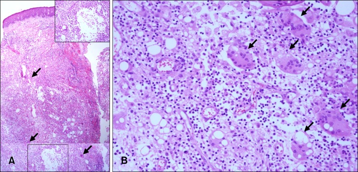
Fig. 2.

Histopathology of skin lesions. (A) An acanthotic epidermis with a marked degeneration of adipocytes, and an intense inflammatory cell infiltrate, composed of lymphocytes and neutrophils. (B) Foreign-body-type giant cells with translucent intracytoplasmic vacuoles (arrows) (H&E; Inset: ×100, A: ×40, B: ×400).
