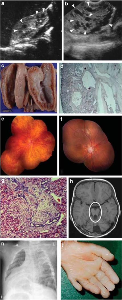
Figure 1. Representative images of degenerative and dysplastic clinical features observed in nephronophthisis (NPHP)-related ciliopathies.
(a) Renal ultrasound from an individual with (degenerative) NPHP. Cysts occur only at the corticomedullary junction and are marked with arrowheads. (b) Renal ultrasound from an individual with (dysplastic) Meckel–Gruber syndrome (MKS, A1633-22). Cysts occur throughout an enlarged kidney and are marked with arrowheads. (c) Autopsy kidney specimen from a fetus with MKS showing bilateral enlarged kidneys interspersed with small, pinhead-sized cysts. (d) Renal histology showing cystic dysplastic kidneys with marked interstitial fibrosis in MKS. (e) Ophthalmoscopy reveals retinitis pigmentosa in the right eye of a patient with (degenerative) Senior–Løken syndrome. (f) Left eye of an NPHP patient with diffuse retinal atrophy with markedly pale discs and enlarged cup. (g) Liver histology showing dysgenesis of the hepatic portal triad with hyperplastic biliary ducts and congenital hepatic fibrosis. (h) Brain magnetic resonance imaging with ‘molar tooth sign’ (marked with a circle) demonstrating vermis hypoplasia, a dysplastic phenotype observed in a JBTS patient. (i) Chest X-ray image of a patient showing situs inversus. (j) Postaxial hexadactyly in an MKS fetus.

