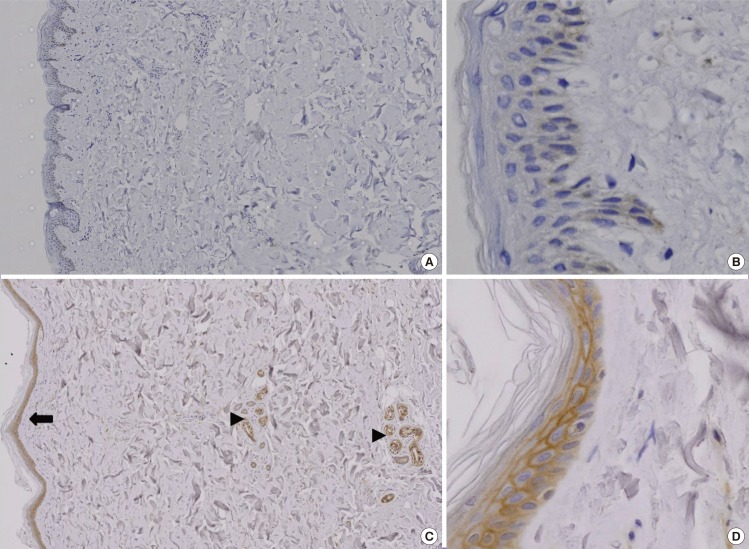
Fig. 1.
Immunohistochemical study of KAI1 protein
The color brown denotes a positive stain. (A) Fewer than 25% of the cells in the normal skin tissue were stained (immunochemical stain, 20×). (B) KAI1 was weakly stained in the normal skin tissue (immunochemical stain, 200×). (C) The diabetic skin tissue displayed increased expression of KAI1. KAI1 immunoreactivity was present in the peripheral membranous staining of keratinocytes (black arrow heads) and appendageal structures (black arrow) (immunochemical stain, 20×). (D) In the diabetic skin tissue samples, KAI1 immunoreactivity was present in the peripheral membrane staining of keratinocytes (immunochemical stain, 200×).

