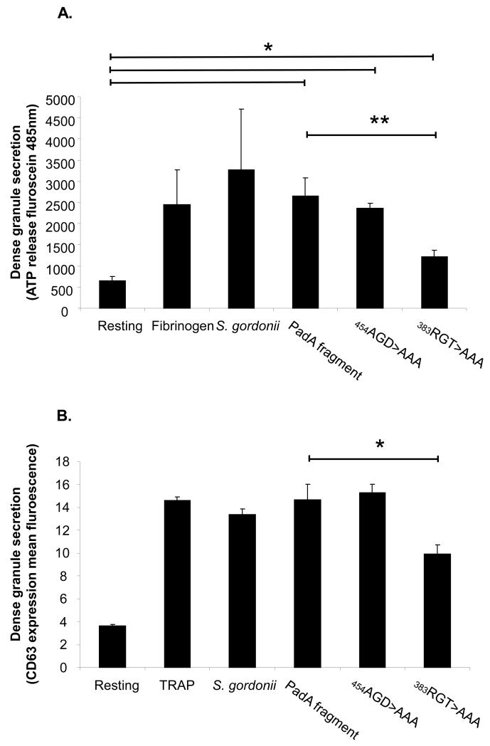
Figure 4. Platelet dense granule secretion following adhesion to Streptococcus gordonii PadA.
(A) Samples were taken from the platelets adhered to immobilized substrates and transferred to a 96 well microtitre plate. The extent of dense granule secretion from the platelets was measured by addition of a chronolume luciferin/luciferase mix. Luminescence was read at 485nm in a microtitre plate reader. (B) Platelets were incubated with S. gordonii for fragments for 10 min. CD63 or appropriately labelled isotype control was added to the platelets. After 20 min incubation the samples were diluted with 1 ml of PBS and analysed on a FACSCalibur flow cytometer. *P<0.05, **P<0.005, n=3-5

