Abstract
Soldiers returning from Iraq have reported respiratory symptoms. Lung biopsies show constrictive bronchiolitis and vascular remodeling with polarizable crystals. We hypothesized that ground surface dust may be a contributing factor to Iraq Afghanistan War Lung Injury (IAW-LI) and analyzed soil grab samples from Camp Victory, Iraq to determine if particle sizes are respirable. Samples contain particles 2.5 micron in size and have sharp edges. Trace metals (including titanium), calcium and silicon are present. Mice with airway instillation of dust have polarizable crystals and septate inflammation. CD4+CD25+FOXP3+ regulatory T cells are decreased in spleen and thymus from mice exposed to dust. IL-2 is elevated from bronchoalveolar lavage taken from dust-exposed mice. Respirable Iraq dust leads to lung inflammation in mice similar to that seen in patients, particularly regarding polarizable crystals which, appear to be titanium.
Keywords: Iraq, Afghanistan, dust, burn pits, lung injury, inflammation, IL-2, regulatory T cells
INTRODUCTION
The U.S. Army Millennium Study determined that 14% of all soldiers in Iraq had new-onset respiratory symptoms. We found a similar finding that 14% of veterans deployed to Iraq and Afghanistan had new-onset respiratory symptoms. Recent reports of bronchiolitis and vascular remodeling among open lung biopsies of symptomatic military personnel supports the concept of inhalational lung injury.
Plausible exposures include inhalation of surface dust, particularly during sandstorms.
Iraq Afghanistan War Lung Injury (IAW-LI) has been described by our group to encompass new-onset respiratory symptoms among previously-healthy young soldiers after deployment to Iraq and Afghanistan.1 The current study was designed to determine if Iraq dust has physico-chemical properties to yield particulate matter air pollution and if inhaled Iraq dust leads to immunosuppression via suppression of regulatory T cells (Treg), with upregulation of pro-inflammatory cytokines.
War-related lung injuries have emerged as previously-unrecognized health problems. Physicians did not recognize the nature of illness in the beginning of the Wars in Iraq and Afghanistan. During the last years of the Iraq War, as well as the ongoing Afghanistan War, new-onset lung problems have been increasingly recognized as being related to deployment in these two countries. Investigators, including our group, have identified asthmatic symptoms or pulmonary function test abnormalities. The Social Security Administration is now compensating for Iraq and Afghanistan War-related lung disease. The Millennium Cohort Study identified 14% of all U.S. soldiers in Iraq with new-onset respiratory symptoms.2
So far, we described a new entity called Iraq-Afghanistan War Lung Injury (IAW-LI), characterized by new, recent-onset respiratory symptoms necessitating spirometry, among 14% of 1,787 Long Island-based Veterans of the Wars in Iraq and Afghanistan.1 Symptoms include dyspnea, exercise-induced shortness of breath, cough, wheezing, chest tightness. Six percent of 1,787 soldiers have spirometric evidence of new airway obstruction, of which half are reversible with bronchodilators; and, the other half has fixed airway obstruction.3

Among the 8% with other new lung diseases are those with: bronchiolitis and pulmonary vascular remodeling—similar to the pathology in lung transplant patients with rejection;5 high levels of iron and titanium in lung tissue in some patients;4 one of our patients has Mycobacterium Avium Complex in lung along with titanium; others have had patterns of granulomatous lung disease, suggestive of either sarcoidosis or hypersensitivity pneumonitis.
Plausible causes of IAW-LI include: Iraq dust, containing sharp particles (<5 µm in size), minerals, metals and sometimes microbes made airborne during sandstorms; “burn pit” fumes from open air burning of trash with jet fuel, JP-8, without incinerators, in large pits; outdoor and indoor aeroallergens;8 improvised explosive devices aerosolizing metals; and blast overpressure, with shock waves related to explosions damaging lung tissue and rendering it vulnerable to further injury.
We seized the opportunity to test if a sample of dust from Camp Victory, Iraq, can induce lung injury with a specific aim to dissect the immunopathogenesis with respect to pro-inflammatory cytokine upregulation and suppression of immune-tolerant regulatory T cells.
MATERIALS AND METHODS
This study evaluated: 1) whether a dust sample from Iraq has physico-chemical properties as particulate matter air pollution; and 2) whether inhaled this Iraq dust in mice induces lung injury via immunosuppresion due to reduced regulatory T cell numbers and increased concentrations of pro-inflammatory cytokines. Iraq dust was analyzed by electron microscopy for particle size, shape, and metal constituents. Particle sizes were 5 microns or less and were sharp but not hollow. Titanium was present in trace amounts. Wild-type C57BL/6 mice were anesthetized and tracheotomized for instillation of 0.1 ml of 1 mg/ml g dust/ 1ml normal saline via tracheostomy tube. A dust sample was collected from Camp Victory, Iraq by the U.S. Army Corps of Engineers and compared to (National Institutes of Standards and Technology) NIST San Joaquin, California inert negative control soil samples and NIST Montana (positive control soil from a titanium mine), in comparison to untreated control mice. All dust-challenged mice had inflammation but the greatest histologic score was in Iraq dust-exposed mice (septate thickening, cellular inflammation, polarizable crystals). Bronchoalveolar lavage cytokine levels were highest for IL-2 in the Iraq dust group and suppression of splenic regulatory T cells CD4+CD25+ was the most in the Iraq dust group.
Dust
The mineral constituents of the dust sample were determined using a Scintag Pad X-ray diffractometer with a Cu Kα radiation (λ = 1.54 Å) from 4 to 90 degrees 2-theta in continuous scan mode at 2 degrees 2-theta/minute. X-ray fluorescence spectroscopy (Bruker S4 Pioneer) was used for elemental analysis. The specific surface area of the dust sample, determined with a QuantachromeNOVA5-point BET analyzer, is 26.429 m2/g. The morphology and size of the dust particles was determined using a scanning electron microscope (SEM; LEO 1550 SFEG) with an energy dispersive X-ray spectrometer (EDS) operating at 15 kV and 30 µA.
Animal Model
Five 8-week-old male C57BL/6 mice served as untreated controls, without exposure to dust. We administered dust 0.1 ml of a 1 mg/ 1 ml sterile water solution intra-tracheally to eight additional age- and gender matched C57BL/6 mice. Mice were anesthetized with ketamine and xylazine prior to insertion of the tracheostomy tube. Mice were examined 1 month after dust exposure. The wild-type (WT) C57BL/6 mice used in this study are from Taconic Laboratories (Germantown, NY). All experiments and animal care procedures are approved by the Institutional Animal Care and Use Committee and conducted according to National Institutes of Health Guide for the Care and Use of Laboratory Animals.
Method: Mice anesthetized with ketamine/xylazine were tracheotomized and then were exposed to 0.01g dust in 0.1 ml normal saline; then the tracheotomy tube was removed, the wound closed with Surgilock®, and mice were kept alive for 4 weeks. At 4 weeks, the mice again were anesthetized with ketamine, xylazine, a tracheotomy tube was inserted, and the lungs were inflation-fixed with formalin for Hematoxylin and Eosin staining. A pathologist blinded to the identities of the samples used the schemata above to score lung injury (crystal, septate thickening, inflammation in terms of lymphocytes per high power field.) To check for crystals, a polarizing light microscope was used.
For additional mice at 4 weeks, the mice again were anesthetized with ketamine, xylazine, a tracheotomy tube was inserted, and the thymus and spleen were removed as previously described (Szema, BMC Immunology) for flow cytometric analysis of CD4+CD25+FOXP3+ T regulatory cells as a percentage of total cells counted in the lymphocyte gate.
Another group of mice at 4 weeks were anesthetized with ketamine, xylazine, a tracheotomy tube was inserted, and bronchoalveolar lavage of 1.5 ml of a solution at a concentration of 1 tablet of Complete Mini antiprotease per 10 ml normal saline was used. Return lavage fluid was stored in an Eppendorf tube dipped in liquid nitrogen and frozen at −80C for analysis at Assaygate Corporation for concentrations in pg/ml of cytokines IL-2, IFN-g, IL-6, and IL-10.
Immunologic Evidence
Our flow cytometric analysis depended on the characteristic surface markers CD25 and CD4, identified by specific antibodies to identify Tregs.7 We also used an additional antibody: FOXP3, an intracellular activation marker, which is critical for Treg survival.9– 14
The spleen and thymus were removed and placed in a Falcon tube with RPMI media and stored on ice until labeling with antibodies for flow cytometric analysis. The spleen and thymus were teased apart and disaggregated on top of a cell strainer in a cell culture dish containing 5 ml of cold media (FACS wash). The cell suspension was transferred to a 50 ml conical tube, centrifuged 10 minutes, resuspended in 5 ml RBC lysis buffer, and incubated for 3 minutes at room temperature. The cells were then centrifuged and washed twice with 15 ml FACS wash buffer, 350 × g 5 minutes and were resuspended at 5 × 10⁶/ml.7
The cells were labeled with 5 microliter One step staining Mouse Treg FlowTM kit (FoxP3 Alexa Fluor® 488/CD25PE/CD4 Per CP) with 100 μ of cell suspension per 5 µl antibody for 20 minutes) followed by fixing with FoxP3 fix/perm buffer (Biolegend Cat. No. 421401) at room temperature for 20 minutes. The cells were then permeabilized in FoxP3 perm buffer (Biolegend Cat No. 421402,) for 20 minutes, centrifuged and washed twice with FoxP3 perm buffer. The cells were then labeled with anti-Helios (22f6) (Biolegend Cat. No. 137203 Alexa Fluor® 647) for 20 minutes at room temperature, centrifuged, washed twice, resuspended, and analyzed by flow cytometry. Lymphocyte and macrophage populations were defined by forward and sideward scatter, according to cell granularity and size. Mean control Treg counts in spleen of 435 was based on our earlier paper with N=10 mice.7
Histological evidence
To test for the presence of airway inflammation, a corollary of airway hyperresponsiveness in asthma, we subject the lungs from mice to tracheostomy under ketamine/xylazine anesthesia and histological examination after inflation fixation in formalin followed by hematoxylin and eosin staining by a pathologist who is blinded to the identities of the samples.6 All abnormalities are graded 1, 2, 3, or 4, based on the intensity and extent of peribronchiolar and perivascular cellular infiltration by a pathologist blinded to the identities of the samples. The pathologist also graded the severity and location of crystal deposition and determined if the crystals, if present, were polarizable.
Experimental Design: Analyses (Collection of Data, Statistical Analysis, and Interpretation)
Results are expressed as mean±S.D. with n being the number of animals per group. For statistical analyses, responses are converted to their logarithms (log10) and differences among groups are analyzed using analyses of variance for repeated measurements (ANOVA) followed by Bonferroni or Tukey’s multiple comparisons with p < 0.05 considered statistically significant.
We seal off the trachea with Vetglue or SurgiLock and examine in 1 month after dust administration.
At 1 month, we will use C02 euthanasia and then measure Treg from thymus and spleen using the methods described in the preliminary studies section, with flow cytometry for markers of Treg (CD4+CD25+FOXP3+).
Histologic sections of H&E-stained lung were graded by a blinded pathologist. We also conduct bronchoalveolar lavage and obtain lung tissue for genomics and proteomics analyses. ANOVA is used to compare Treg numbers among the four groups of dust-treated animals, compared to untreated controls.
Lung cytokines
As added evidence of possible airway inflammation, we perform bronchoalveolar lavage (BAL) in WT mice. The lungs of each mouse are lavaged three times with 1 ml of PBS, including an EDTA-free protease inhibitor cocktail (Roche Diagnostics, Indianapolis, IN). The BAL fluid is centrifuged at 400 g for 5 min at 21°C, and the supernatant is analyzed by a quantitative ELISA assay (Assaygate) for selected inflammatory cytokines, IL-2, IL-4, IFN-g, IL-10.
RESULTS
Dust
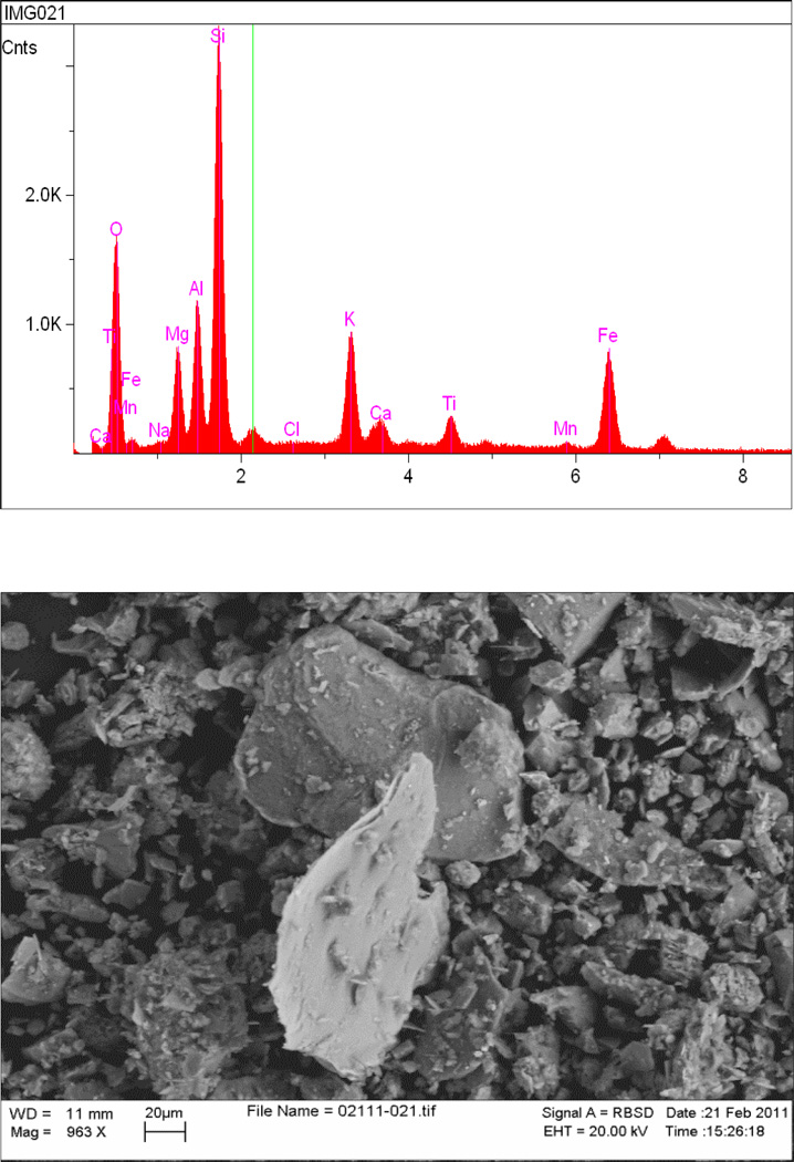
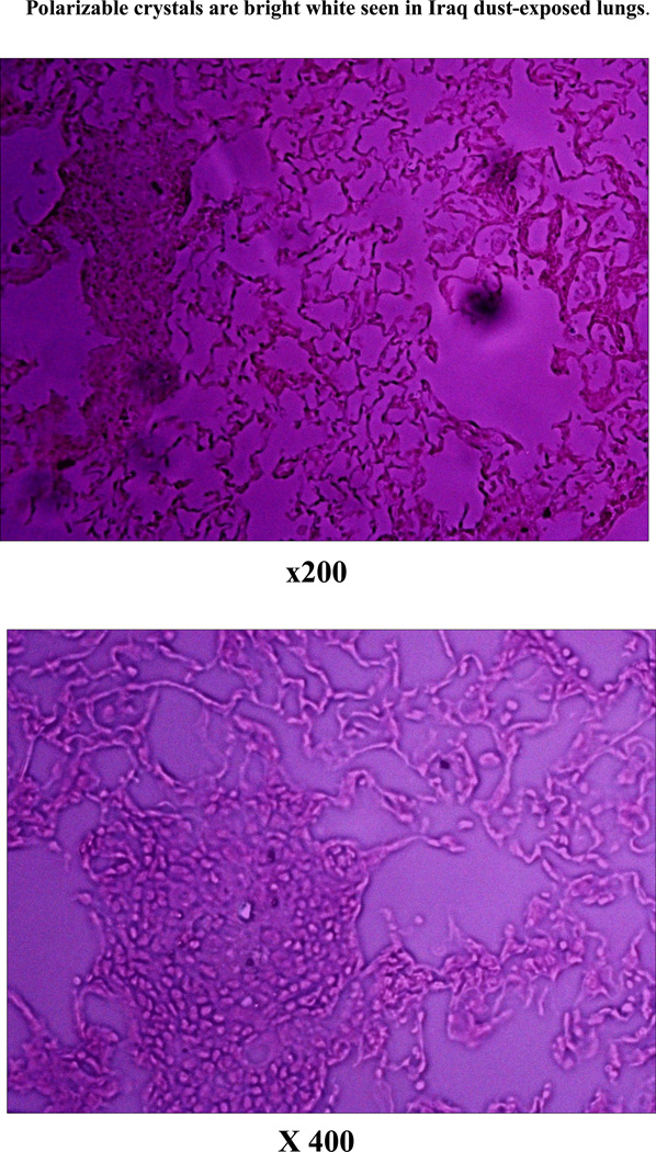
Particle sizes of 5 microns or less are present in the Iraq dust sample. The particles are angular in shape, with sharp edges, but are not hollow. Titanium, iron, silicon, calcium and other metals, are present in Iraq dust as shown by energy-dispersive x-ray analysis (Figure 1). Histologic airway inflammation and interstitial inflammation are not present in untreated C57BL/6 mice but are greatest in Iraq dust-exposed mice compared to San Joaquin or Montana dust experimental groups. The Iraq dust-treated group had the most crystals (which were polarizable), septate thickening, and inflammation. The San Joaquin dust generated less inflammation as a negative control compared to Montana dust (laden with titanium) as a positive control. The overall increase in the triad of crystals, septate thickening, and inflammation in the Iraq dust exposed mice suggests that titanium alone, while deleterious, does not account for the entire pathophysiologic process.
Figure 1). Dust.
Energy-dispersive X-ray analysis of representative particle in Iraq dust sample, indicating elements detected (top). Electron micrograph of Camp Victory, Iraq, dust sample shows variable particle sizes, including fraction less than 5µm (respirable). Particles are angular with sharp edges (bottom).
The immunopathogenesis of Iraq dust lung injury supports suppression of activated Treg (CD4+CD25+FOXP3+) cells which are immune-tolerant lymphocytes. These are anti-inflammatory. Suppression of total Treg (CD4+CD25+) is associated in these experiments with upregulation of pro-inflammatory cytokine IL-2. In contrast, IL-4, which skews B cells to produce the allergic antibody IgE, is not increased. The Montana dust, titanium-laden, strongly upregulated anti-inflammatory cytokine IL-10 uniquely among the groups.
Animal Model
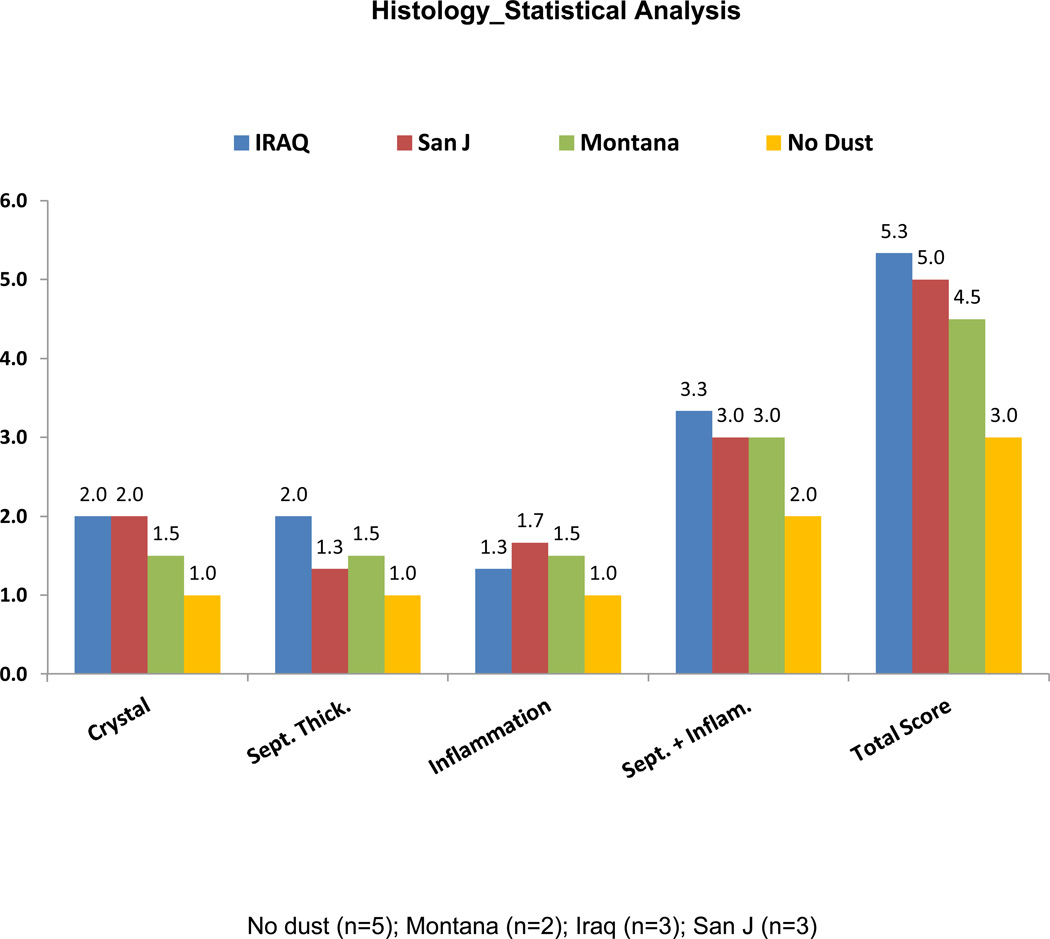
Figure 2: Histology Scores Among Lungs from Mice Exposed to dust from Iraq, California or Montana
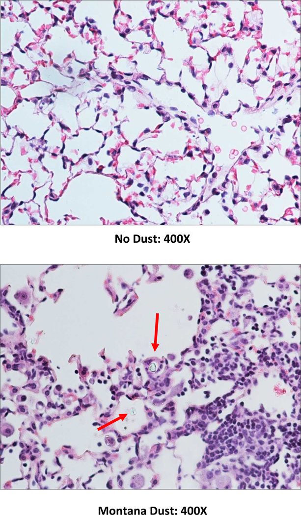
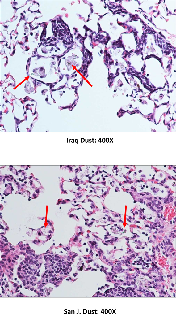
Figure 2). Histology.
Histologic examples of lungs from each experimetnal arm shows: no dust at 400× with normal histology in C57BL/6 mouse lung; San Joaquin, California dust with a minimal amount of focal lymphocytic accumulation in the lower right hand corner of the field; Montana dust with significant inflammation, more widespread; and Iraq dust with septate thickening, interstitial inflammation, incompletely phagocytosed crystals.
No dust (n=5); Montana (n=2); Iraq (n=3); San J (n=3)
Iraq-exposed mice had the most lung injury, based on a scoring system utilizing: 1) presence of crystals, 2) septate thickening, and 3) inflammation.
A pathologist blinded to the identities of treated mice scored for the presence of crystals, septate thickening, or inflammation, using a grading scheme for 1=no crystal, 2= few crystals, and 3=numerous crystals; and 1=no septate thickening vs. 2=septate thickening present. A total score added the presence of all three characteristics. Crystals were identified by birefringence in polarizing light microscopy.
Three mice exposed to Iraq dust had the highest mean scores or 5.3, vs. 5.0 for San Juan, 4.5 for Montana, and 3.0 for no dust. The presence of titanium in Montana dust but lower score thresholds suggests that titanium alone is not sufficient to induce the combined presence of numerous crystals, septet thickening, and inflammation.
Immunologic evidence
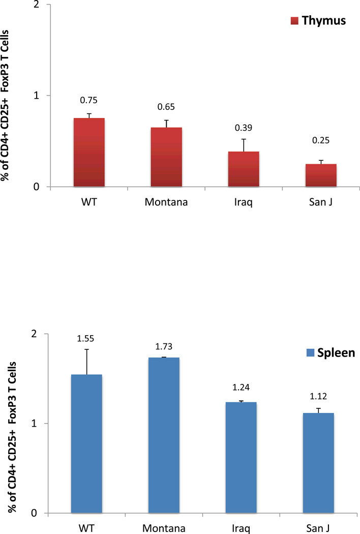
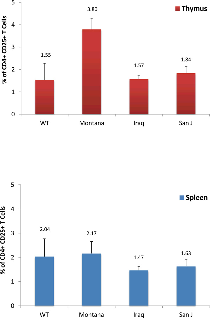
Figure 3: Flow Cytometric Scores from thymus and spleen of dust-exposed mice
Figure 3). Flow cytometry.
CD+CD25+ Treg cell percentages were lowest in spleen in Iraq-exposed mice compared to all other groups. Activated CD4+CD25+FOXP3+ Treg percentages in Iraq-exposed mice were the second lowest among all groups for both thymus and spleen.
Regulatory T cells (Treg) as defined by CD4+CD25+T cell percentages of total cells counted from thymus were lowest among the mice exposed to Iraq dust, suggesting the most immunosuppression. Treg are immune-tolerant cells; loss of Treg suggests lack of immune tolerance. In contrast Treg were highest in mice exposed to dust from Montana. Even though Treg in Iraq-exposed lungs were lowest among the 3 dust-treated groups, this number was similar to wild-type mice. Since the thymus is the central origin of Treg, a surrogate may be the spleen, since peripheral immune tolerance occurs in the latter organ.
Splenic Treg were lowest in the Iraq-exposed mice versus all other groups, including wild-type, Montana, and San Joaquin, California dust. These data indicate suppression of immune-tolerant CD4+CD25+ regulatory T cell numbers in the periphery.
Triple-staining CD4+CD25+FOXP3+ Treg indicate activation and survival. Percentages of these CD4+C25+FOXP3+ cells were all lower in dust-treated groups, compared to untreated mice. In descending order of percentages: Montana, followed by Iraq, then San Joaquin, California in thymus.
In the spleen, numbers of triple-staining, CD4+CD25+FOXP3+ Treg were lower in Iraq and San Joaquin-exposed mice, compared to wild-type and Montana experimental arms.
Lung cytokines
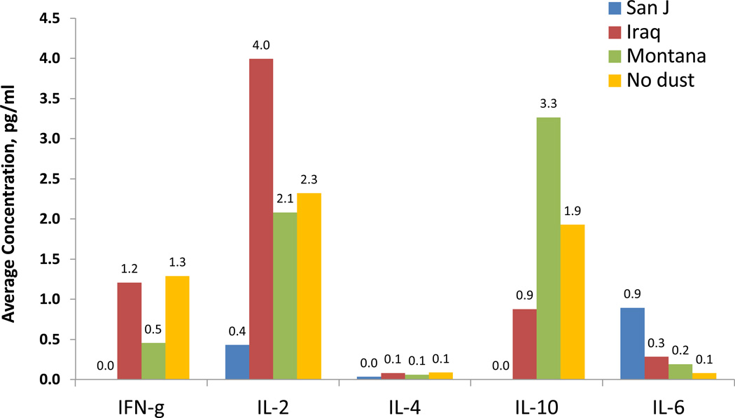
Figure 4: Cytokine concentrations in bronchoalveolar lavage fluid from mice exposed to either of the 3 dusts
Figure 4). Cytokines analysis.
Levels of cytokines were highest for IL-2 in the Iraq group, supporting a pro-inflammatory milieu with Iraq dust exposed, compared to San Joaquin, Montana, and untreated mice.
IL-4 was nearly undetectable, thus supporting the concept that this exposure does not induce an IgE-mediated allergic response. Interferon gamma levels were less pronounced than IL-2 but were highest in Iraq and untreated groups, possibly induced as a counter-regulatory response. Anti-inflammatory IL-10 was lowest in the Iraq group among the dust, with Montana and no dust having the highest response. IL-6 was higher in San Joaquin compared to the other groups, which were negligible.
DISCUSSION
Particulate matter air pollution induces vascular inflammation and is associated with premature death from cardiovascular and lung disease, including myocardial infarction and asthma exacerbations.15, 16
In the case of particulate matter air pollution from an Iraq dust sample, we now know that dust size is respirable, with 2.5 micron-sized particles present, and that these particles exhibit sharp edges. The physical properties are a concern, akin to asbestos fibers, as the histological slides show incomplete phagocytosis by pulmonary alveolar macrophages. A plausible theory as to shape is that heavy trucks and tanks may have crushed surface dust to alter its rheology. These are solid particles. We did not see hollow particles discussed by Captain Mark Lyles at the 1st Scientific Symposium on Lung Health After Deployment to Iraq and Afghanistan, Stony Brook University, March, 2011(personal communication, Captain Mark Lyles, U.S. Naval War College, Newport, RI). So, these particles cannot transport substances as containers or “nano-carrier vehicles.” Concentrations of trace metals and minerals in the dust are concerning, since some forms of titanium may be pro-fibrotic17 and is not digestable, and calcium is an airway irritant.
Instillation of Iraq dust yields lymphocytic airway inflammation and interstitial changes which are patchy. Subtle changes in lungs of patients with dust inhalation may not necessarily be visualized with chest x-rays or observed with in-office spirometry, as seen in the cohort of patients studied by Matt King and Robert Miller’s group.18 These patients have lungs which reflect the concept that histologic bronchiolitis and vascular remodeling may be present in the absence of radiographic and pulmonary function physiologic abnormalities. The polarizable crystals we see in mice with dust inhalation are similar to that reported by Miller’s group. Among our three patients we have examined by open lung biopsy, all have crystals, and titanium is the suspected culprit by micro x-ray fluorescence.
Interleukin 2 or IL-2 is a pro-inflammatory cytokine expressed by lymphocytes which enhances further lymphocyte proliferation. The striking finding of higher levels of IL-2 in Iraq dust-treated mice supports a pro-inflammatory milieu, especially since anti-inflammatory IL-10 levels are not upregulated. This is a specific inflammation profile, since IL-6, another pro-inflammatory cytokine, notably increased in Acute Respiratory Distress Syndrome from lung stretch,19 is not increased. IFN-g is also not increased.
Regulatory T cells or Treg are immune-tolerant cells which are anti-inflammatory. They are centrally located in the thymus and also may be found in the periphery from a splenic origin. Finding suppressed Treg staining for CD4+CD25+ surface markers suggests that there is peripheral immunosuppression in dust-treated mice.
While a plethora of airborne hazards may have been experienced by military personnel during deployment to Iraq—detonation of mortars by Al-Qaeda, improvised explosive devices, vehicle improvised explosive devices, blast overpressure from shock waves resulting from explosions, dust and sandstorms called Shamal and Sharq, fumes from burning trash with jet fuel JP-8 in continuously incendiary burn pits, indoor and outdoor aeroallergens (depending on geographic location), infectious vectors from organic components in the dust and air—both bacterial and viral and fungal—our model allows the isolation of dust and the ability to examine its sundry inorganic components and their effect on lung health.
Since only half of patients with new-onset dyspnea after deployment to Iraq and Afghanistan in our cohort1 respond to asthma medications, targeting the specific, characteristic immune response with respect to Treg and IL-2 may be a more focused approach.
While this research provides some insight into the role of dust encountered in Iraq and Afghanistan Wars on the production of respiratory disease observed in some of the troops deployed there, more work is needed to understand the role of other airborne contaminants in producing these effects. Additionally, the study results are based on only dust samples obtained at Camp Victory, Iraq. Furthermore, the number of animals tested was relatively small. Holding some animals beyond one month to evaluate long-term effects would be of interest.
TABLE 1.
P values based on ttest
| Crystal | Septate Thickening | Inflammation | Septate + Inflammation | Total Score | |
|---|---|---|---|---|---|
| WT/Mont | 0.117 | 0.117 | 0.117 | 0.117 | 0.117 |
| WT/Iraq | 0.000 | 0.000 | 0.220 | 0.002 | 0.000 |
| WT/Sanj | 0.055 | 0.220 | 0.034 | 0.055 | 0.055 |
| Mont/Iraq | 0.272 | 0.272 | 0.789 | 0.724 | 0.537 |
| Mont/Sanj | 0.591 | 0.789 | 0.789 | 1.000 | 0.806 |
| Iraq/Sanj | 1.000 | 0.116 | 0.519 | 0.643 | 0.795 |
The highest total lung injury score was in the Iraq dust-exposed group, with more crystals, which were polarizable, consistent septate thickening, and inflammation, both airway and interstitial.
Flow cytometry_Statistical Analysis
| ANOVA | ||||||
|---|---|---|---|---|---|---|
| Sum of Squares | df | Mean Square | F | Sig. | ||
| Between Groups | 11.300 | 3 | 3.767 | 2.820 | .099 | |
| Thymus_CD4CD25 | Within Groups | 12.020 | 9 | 1.336 | ||
| Total | 23.320 | 12 | ||||
| Between Groups | .957 | 3 | .319 | 2.211 | .156 | |
| Spleen_CD4CD26 | Within Groups | 1.298 | 9 | .144 | ||
| Total | 2.255 | 12 | ||||
| Between Groups | .576 | 3 | .192 | 9.452 | .004 | |
| Thymus_CD4CD25_FoxP3 | Within Groups | .183 | 9 | .020 | ||
| Total | .759 | 12 | ||||
| Between Groups | .652 | 3 | .217 | 1.233 | .353 | |
| Spleen_CD4CD25_FoxP3 | Within Groups | 1.586 | 9 | .176 | ||
| Total | 2.237 | 12 | ||||
| 1= No dust (n=5); 2= Montana (n=2); 3= Iraq (n=3); 4= San J (n=3) | ||||||
| P value-Based on Tukey Post Hoc Test | ||||
|---|---|---|---|---|
| Thymus_CD4CD25_FoxP3 | Tukey HSD | 1 | 2 | 0.819 |
| 3 | 0.027 | |||
| 4 | 0.004 | |||
| 2 | 1 | 0.819 | ||
| 3 | 0.248 | |||
| 4 | 0.054 | |||
| 3 | 1 | 0.027 | ||
| 2 | 0.248 | |||
| 4 | 0.656 | |||
| 4 | 1 | 0.004 | ||
| 2 | 0.054 | |||
| 3 | 0.656 | |||
| P value based on unpaired ttest |
|---|
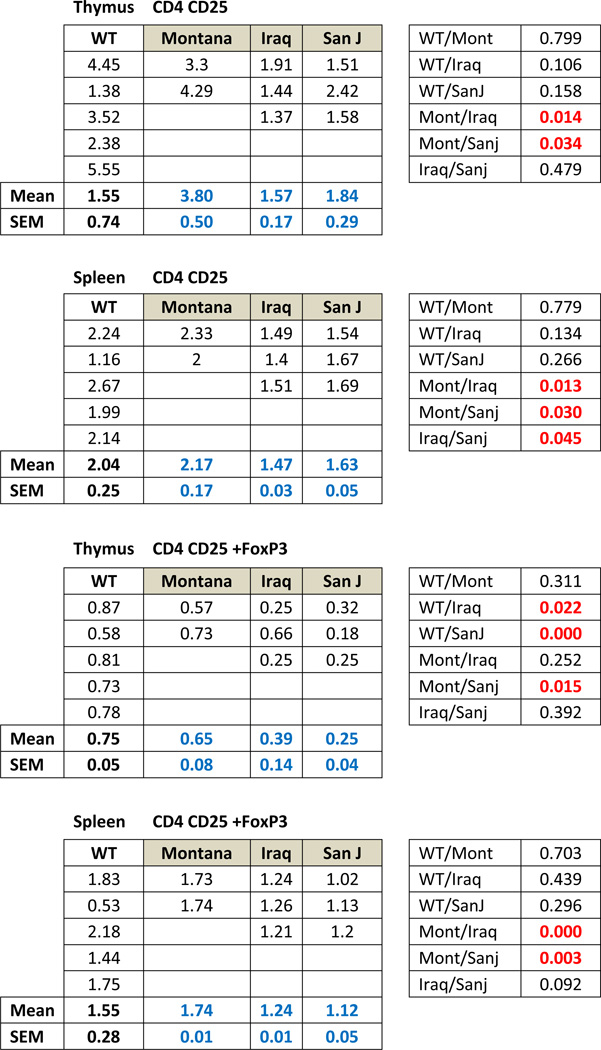 |
TABLE 3.
BAL Cytokines_Statistical Analysis ANOVA
| ANOVA | ||||||
|---|---|---|---|---|---|---|
| Sum of Squares | df | Mean Square | F | Sig. | ||
| Between Groups | 3.867 | 3 | 1.289 | .832 | .509 | |
| IFNg | Within Groups | 13.945 | 9 | 1.549 | ||
| Total | 17.812 | 12 | ||||
| Between Groups | 19.174 | 3 | 6.391 | 3.203 | .076 | |
| IL2 | Within Groups | 17.959 | 9 | 1.995 | ||
| Total | 37.132 | 12 | ||||
| Between Groups | .004 | 3 | .001 | .287 | .834 | |
| IL4 | Within Groups | .046 | 9 | .005 | ||
| Total | .051 | 12 | ||||
| Between Groups | 14.911 | 3 | 4.970 | 1.697 | .237 | |
| IL10 | Within Groups | 26.360 | 9 | 2.929 | ||
| Total | 41.271 | 12 | ||||
| Between Groups | 1.323 | 3 | .441 | .780 | .534 | |
| IL6 | Within Groups | 5.088 | 9 | .565 | ||
| Total | 6.411 | 12 | ||||
| 1= No dust (n=5); 2= Montana (n=2); 3= Iraq (n=3); 4= San J (n=3) | ||||||
| P value based on unpaired ttest | |||||
|---|---|---|---|---|---|
| IFN-g | IL-2 | IL-4 | IL-10 | IL-6 | |
| WT/Mont | 0.569 | 0.708 | 0.738 | 0.515 | 0.527 |
| WT/Iraq | 0.942 | 0.236 | 0.924 | 0.487 | 0.106 |
| WT/Sanj | 0.275 | 0.006 | 0.401 | 0.221 | 0.263 |
| Mont/Iraq | 0.211 | 0.433 | 0.789 | 0.106 | 0.579 |
| Mont/Sanj | 0.272 | 0.011 | 0.696 | 0.047 | 0.588 |
| Iraq/Sanj | 0.009 | 0.093 | 0.421 | 0.017 | 0.533 |
CLINICAL RELEVANCE.
Surface dust from Iraq is respirable particulate matter air pollution, with physico-chemical properties leading to inflammation and immunosuppression in mice. This suggests inhaled dust may contribute to development of new-onset dyspnea among soldiers.
Footnotes
- Recently completed grant from Merck to study dust mite antigen on lung epithelial cells in vitro $15K Sept 2013
- Garnett McKeen Labs completed grant July 2012 to study drug RUX as an anti lung injury agent for Iraq dust $60K
- NIEHS R21 2013-6 co investigator to study asthma in Hurricane Sandy victims who were previously World Trade Center disaster workers (PI Adam Gonzalez, PhD, Stony Brook University)
- Grant applications submitted to VA Merit Review Program to study regulatory T cells in mice exposed to Iraq dust fall 2013
- Grant application submitted to Lupus Research Institute to study regulatory T cells in VIP knockout mice December 2013
- In contract negotiation with Phasebio Corporation as a consultant for their VIP drug December 2013
- Developing a smartshirt to measure lung physiology with associate dean of curriculum at Stony Brook University and associate Dean of Engineering December 2013
- Private practice Three Village Allergy & Asthma, PLLC, South Setauket, NY
- Chief, Allergy Section, Veterans Affairs Medical Center, Northport, NY
Contributor Information
Anthony M. Szema, Email: anthony.szema@stonybrookmedicine.edu.
Richard J. Reeder, Email: rjreeder@stonybrook.edu.
Andrea D. Harrington, Email: harrinan@gmail.com.
Millicent Schmidt, Email: millicentpschmidt@gmail.com.
Jingxuan Liu, Email: jingxuan.liu@stonybrookmedicine.edu.
Marc Golightly, Email: marc.golightly@stonybrookmedicine.edu.
Todd Rueb, Email: todd.rueb@stonybrook.edu.
Sayyed A. Hamidi, Email: hhamidi55@hotmail.com.
BIBLIOGRAPHY
- 1.Szema AM, Salihi W, Savary K, Chen JJ. Respiratory symptoms necessitating spirometry among soldiers with Iraq/Afghanistan war lung injury. Journal of Occupational and Environmental Medicine. 2011;53:961–965. doi: 10.1097/JOM.0b013e31822c9f05. [DOI] [PubMed] [Google Scholar]
- 2.Smith B. Newly reported respiratory symptoms and conditions among military personnel deployed to Iraq and Afghanistan: a prospective population-based study. Am J Epidemiol. 2009 Dec 1;170(11):1433–1442. doi: 10.1093/aje/kwp287. [DOI] [PubMed] [Google Scholar]
- 3.Szema AM, Peters MC, Weissinger KM, Gagliano CA, Chen JJ. New-onset asthma among soldiers serving in Iraq and Afghanistan. Allergy and Asthma Proceedings : the official journal of regional and state allergy societies. 2010;31:67–71. doi: 10.2500/aap.2010.31.3383. [DOI] [PubMed] [Google Scholar]
- 4.Szema AM, Schmidt MP, Lanzirotti A, et al. Titanium and iron in lung of a soldier with nonspecific interstitial pneumonitis and bronchiolitis after returning from Iraq. Journal of Occupational and Environmental Medicine. 2012;54:1–2. doi: 10.1097/JOM.0b013e31824327ca. [DOI] [PubMed] [Google Scholar]
- 5.King MS, Eisenberg R, Newman JH, et al. Constrictive bronchiolitis in soldiers returning from Iraq and Afghanistan. The New England Journal of Medicine. 2011;365:222–230. doi: 10.1056/NEJMoa1101388. [DOI] [PMC free article] [PubMed] [Google Scholar]
- 6.Szema AM, Hamidi SA, Lyubsky S, et al. Mice lacking the VIP gene show airway hyperresponsiveness and airway inflammation, partially reversible by VIP. American Journal of Physiology Lung Cellular and Molecular Physiology. 2006;291:L880–L886. doi: 10.1152/ajplung.00499.2005. [DOI] [PubMed] [Google Scholar]
- 7.Szema AM, Hamidi SA, Golightly MG, Rueb TP, Chen JJ. VIP Regulates the Development & Proliferation of Treg in vivo in spleen. Allergy, Asthma, and Clinical Immunology : official journal of the Canadian Society of Allergy and Clinical Immunology. 2011;7:19. doi: 10.1186/1710-1492-7-19. [DOI] [PMC free article] [PubMed] [Google Scholar]
- 8.Szema AM. Climate change, allergies, and asthma. Journal of Occupational and Environmental Medicine. 2011;53:1353–1354. doi: 10.1097/JOM.0b013e318237a00d. [DOI] [PubMed] [Google Scholar]
- 9.Venigalla RK, Tretter T, Krienke S, et al. Reduced CD4+,CD25− T cell sensitivity to the suppressive function of CD4+,CD25high,CD127 −/low regulatory T cells in patients with active systemic lupus erythematosus. Arthritis and Rheumatism. 2008;58:2120–2130. doi: 10.1002/art.23556. [DOI] [PubMed] [Google Scholar]
- 10.Fan H, Cao P, Game DS, Dazzi F, Liu Z, Jiang S. Regulatory T cell therapy for the induction of clinical organ transplantation tolerance. Seminars in Immunology. 2011;23:453–461. doi: 10.1016/j.smim.2011.08.012. [DOI] [PubMed] [Google Scholar]
- 11.Mamessier E, Birnbaum J, Dupuy P, Vervloet D, Magnan A. Ultra-rush venom immunotherapy induces differential T cell activation and regulatory patterns according to the severity of allergy. Clinical and Experimental Allergy. 2006;36:704–713. doi: 10.1111/j.1365-2222.2006.02487.x. [DOI] [PubMed] [Google Scholar]
- 12.Hori S, Nomura T, Sakaguchi S. Control of regulatory T cell development by the transcription factor Foxp3. Science. 2003;299:1057–1061. [PubMed] [Google Scholar]
- 13.Ono M, Yaguchi H, Ohkura N, et al. Foxp3 controls regulatory T-cell function by interacting with AML1/Runx1. Nature. 2007;446:685–689. doi: 10.1038/nature05673. [DOI] [PubMed] [Google Scholar]
- 14.Fontenot JD, Gavin MA, Rudensky AY. Foxp3 programs the development and function of CD4+CD25+ regulatory T cells. Nature Immunology. 2003;4:330–336. doi: 10.1038/ni904. [DOI] [PubMed] [Google Scholar]
- 15.Brook RD, et al. AHA Scientific Statement: Particulate Matter Air Pollution and Cardiovascular Disease. Circulation. 2010;121:2331–2378. doi: 10.1161/CIR.0b013e3181dbece1. [DOI] [PubMed] [Google Scholar]
- 16.Karatksani A, et al. Particulate matter air pollution and respiratory symptoms in individuals having either asthma or chronic obstructive pulmonary disease: a European multicentre panel study. Environ Health. 2012;11:75. doi: 10.1186/1476-069X-11-75. [DOI] [PMC free article] [PubMed] [Google Scholar]
- 17.Sager T, Kommineni C, Castranova V. Pulmonary response to intratracheal instillation of ultrafine versus fine titanium dioxide: role of particle surface area. Part Fibre Toxicol. 2008;5:17. doi: 10.1186/1743-8977-5-17. [DOI] [PMC free article] [PubMed] [Google Scholar]
- 18.King Matthew S, Eisenberg Rosana, Newman John H, Tolle James J, Harrell Frank E, Jr, Nian Hui, Ninan Mathew, Lambright Eric S, Sheller James R, Johnson Joyce E, Miller Robert F. Constrictive Bronchiolitis in Soldiers Returning from Iraq and Afghanistan. N Engl J Med. 2011;365:222–230. doi: 10.1056/NEJMoa1101388. [DOI] [PMC free article] [PubMed] [Google Scholar]
- 19.Gurkan Ozlem U, MD, He Chaoxia, MS, Zielinski Rachel, BS, Rabb Hamid, MD, King Landon S, MD, Dodd-o Jeffrey M, MD, D’Alessio Franco R, Aggarwal Neil, Pearse David, Becker Patrice M., MD Interleukin-6 mediates pulmonary vascular permeability in a two-hit model of ventilator-associated lung injury. Exp Lung Res. 2011 Dec;37(10):575–584. doi: 10.3109/01902148.2011.620680. [DOI] [PMC free article] [PubMed] [Google Scholar]


