SUMMARY
Bacteriophage lambda capsids provide a flexible molecular scaffold that can be engineered to display a wide range of exogenous proteins, including full-length viral glycoproteins produced in eukaryotic cells. One application for such particles lies in the detection of virus-specific antibodies, since they may obviate the need to work with infectious stocks of highly pathogenic or emerging viruses that can pose significant biosafety and biocontainment challenges. Bacteriophage lambda capsids were produced that displayed an insect-cell derived, recombinant H5 influenza virus hemagglutinin (HA) on their surface. The particles agglutinated red blood cells efficiently, in a manner that could be blocked using H5 HA-specific monoclonal antibodies. The particles were then used to develop a modified hemagglutinination-inhibition (HAI) assay, which successfully identified human sera with H5 HA-specific HAI activity. These results demonstrate the utility of HA-displaying bacteriophage capsids for the detection of influenza virus-specific HAI antibodies.
Keywords: Influenza virus, hemagglutinin, H5 HA, hemagglutination inhibition, bacteriophage, lambda
Bacteriophage lambda capsids can provide a highly flexible scaffold for the high density display of foreign peptides and proteins (Gupta et al., 2003; Mikawa, Maruyama, and Brenner, 1996; Santini et al., 1998; Sternberg and Hoess, 1995; Yang et al., 2000). One commonly used method is to fuse the protein or peptide of interest to the major phage capsid protein, gpD, thereby producing phage particles that incorporate the desired protein into their capsids when propagated in E. coli host cells (Mikawa et al., 1996; Santini et al., 1998; Sternberg and Hoess, 1995). However, this approach is suboptimal for glycosylated proteins such as the HIV-1 envelope glycoprotein or the influenza virus hemagglutinin (HA). Consequently, an alternative display approach has therefore been developed which allows one to “decorate” preformed, gpD-deficient phage with exogenously supplied gpD or recombinant gpD-fusion proteins, produced in eukaryotic cells (Mattiacio et al., 2011; Sternberg and Hoess, 1995). This approach permits the decoration of lambda phage capsids with glycoproteins produced in eukaryotic cells, such as the HIV-1 envelope protein (Mattiacio et al., 2011).
To display the influenza virus HA on the surface of lambda phage, a soluble, recombinant gpD:HA protein was produced in insect cells, since these cells have been shown to support the production of biologically functional, glycosylated HA (Treanor, 2009). For this purpose, the HA from a well-characterized H5N1 influenza virus (A/Vietnam/1203/04) was used, and the gpD:H5HA fusion protein was then purified using a C-terminal hexahistidine tag and a nickel-affinity column (Figure S1).
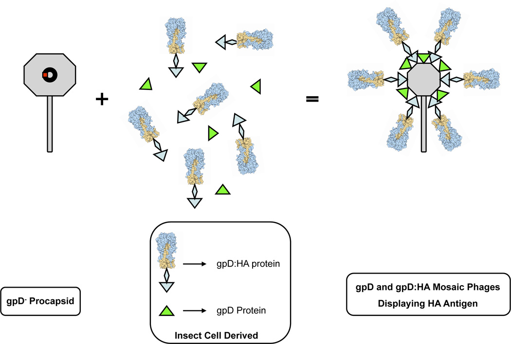
Conditions for the decoration of gpD-deficient phage capsids with the purified, recombinant gpD:H5HA protein were optimized. Attempts to decorate gpD-deficient phage particles with gpD:H5HA protein alone resulted in phage capsids that were unstable in the presence of EDTA, reflecting incomplete occupancy of available gpD binding sites (Figure S2). This is presumably because the large gpD:H5HA protein is sterically hindered from binding all 420 of the gpD binding sites on the capsid (Yang et al., 2000), much like the large gpD:HIV-Env fusion proteins described previously (Mattiacio et al., 2011). To address this problem, “mosaic” phage particles were produced by decorating gpD-deficient capsids with a combination of both gpD:H5HA and wild-type gpD (Mattiacio et al., 2011). This resulted in phage capsids that were stable in the absence of EDTA, which is indicative of full occupancy of the available gpD binding sites on the capsid (Figure S2). A large batch of these “mosaic”, decorated particles was then prepared, purified by CsCl density gradient purification, and analyzed by immunoblot assay using a polyclonal anti-gpD antiserum. This confirmed that both the wild-type and the gpD:H5HA fusion protein were incorporated into the lambda phage capsid (Figure S3). The physical structure of these mosaic, decorated capsids is shown schematically in Figure 1.
Figure 1. Production of lambda phage capsids decorated with HA.
Phage capsids were decorated with HA by adding a mixture of gpD:H5HA fusion protein and wild-type gpD protein to gpD-deficient lambda phage particles. This produced mosaic phage capsids displaying H5 HA at high copy number. Not drawn to scale.
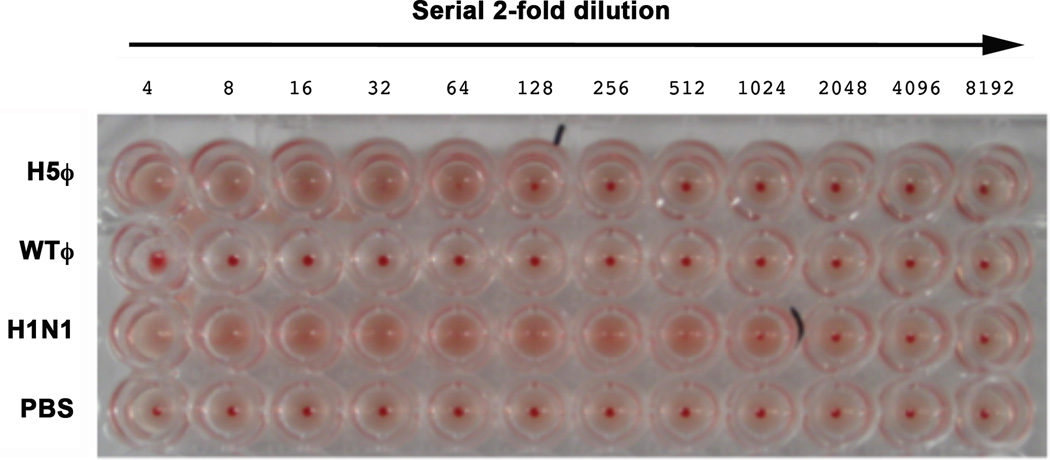
Phage capsids decorated with gpD:H5HA should display HA on their surface in a multivalent array, allowing them to efficiently bind to sialic acid. An experiment was therefore conducted to determine whether phage decorated with recombinant H5 HA could agglutinate chicken red blood cells (cRBC). To do this, cRBC were incubated with serially diluted aliquots of H5HA-displaying phage particles, WT phage particles, a positive control H1N1 influenza virus (A/New Caledonia/20/99) or PBS. H5HA-displaying phage particles agglutinated cRBCs readily when added at concentrations > 5 × 10⁸ PFU/well (Figure 2). The positive control H1N1 influenza virus also agglutinated the cRBCs to a dilution of 1:1024 (consistent with the titer of this virus stock), while WT phage particles and PBS alone did not agglutinate cRBCs (Figure 2).
Figure 2. Phage particles decorated with H5 HA efficiently agglutinate RBCs.
Phage particles (either decorated with H5 HA or not; respectively, H5ϕ and WTϕ), or influenza virus virions (H1N1) were serially diluted in PBS in a 96-well V-bottom plate (Costar); negative control wells contained PBS alone (PBS). 50 µl of a suspension of chicken red blood cells (RBCs) was then added to each well, and samples were incubated at 4°C for 1 hour, after which the plate was photographed. Agglutination titer was determined as the last dilution at which agglutination was detected.
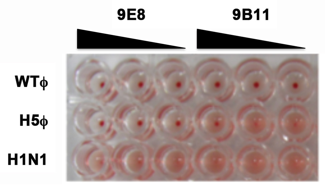
Two H5 HA-specific monoclonal antibodies (Mabs) (kindly provided by Dr. Gary Nabel, NIH VRC) were next used to determine whether HA-specific antibodies could block the hemagglutination activity of H5 HA-displaying phage particles. Mab 9E8 has high H5 specific neutralizing activity, while Mab 9B11 has lower H5-specific neutralizing activity (Yang et al., 2007). Mab 9E8 blocked hemagglutination by H5 HA-displaying phage particles at concentrations as low as 1 ng/well, but had no effect on hemagglutination by H1N1 influenza virus (A/New Caledonia/20/99) (Figure 3). Mab 9B11 also inhibited hemagglutination by the H5 HA-displaying phage, though only at higher concentrations (100 ng/well) (Figure 3).
Figure 3. H5 HA-specific monoclonal antibodies block hemagglutination by phage particles decorated with H5 HA.
Phage particles (H5ϕ or WTϕ), or influenza virus virions (H1N1) were incubated with 100ng, 40ng, or 1ng of Mab 9E8 or 9B11 for one hour at room temperature. Hemagglutination was then tested, using chicken RBC.
These findings suggested the H5 HA-displaying phage particles could have utility in hemagglutination-inhibition (HAI) assays, which are widely used in seroepidemiologic studies of influenzavirus infection, and for the detection of hemagglutinin (HA)-specific antibodies in vaccine trials (Hirst, 1942; Katz, Hancock, and Xu, 2011). The HAI assay typically uses wild-type influenza viruses or HA-pseudotyped viruses matching the antibodies of interest. In the case of wild-type viruses, this can pose challenges in terms of biosafety and biocontainment (Imperiale and Hanna, 2012; Lipsitch and Bloom, 2012); for pseudotyped viruses, a limitation is that such viruses must propagated in specialized cell lines expressing the HA of interest. It was therefore important to test whether HA-displaying phage particles could be used to conduct HAI assays.
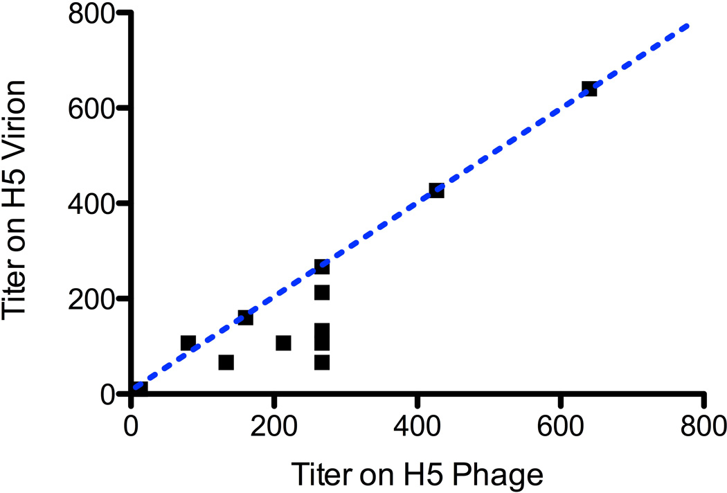
Blinded serum samples from human subjects who participated in a H5 influenza vaccine trial (Keitel et al., 2008; Treanor et al., 2006) were provided, and tested for their ability to inhibit hemagglutination by either H5 HA-displaying phage particles or influenza virions displaying the same H5 HA (for this purpose, a pseudotyped single-cycle infectious Influenza A virus [sciIAV] displaying the HA from the A/Vietnam/03/2004 H5N1 virus was used; (Baker et al., 2013; Martinez-Sobrido et al., 2010)). HAI titers were determined by serial two-fold dilution of test sera, using both phage particles and influenza virions [sciIAV] (Figure 4). The results show that HAI titers measured using phage particles and influenza virions were very nearly identical (Figure 4). The concordance correlation coefficient of the two assays was 0.894 (95% confidence interval: 0.741–0.959); hypothetical perfect concordance is represented by the dotted blue line in Figure 4. Within the 19 samples tested, both assays correctly identified both positive (n = 13; titers ≥ 40) and negative (n = 6; titers < 40) sera, as defined by previous assays using H5N1 IAV. Interestingly, in those instances when the two assays yielded slightly different results, the phage-based assay appeared to be more sensitive (i.e., gave modestly greater titers).
Figure 4. Phage particles decorated with H5 HA can be used to detect HAI antibodies in human sera.
Phage particles decorated with H5 HA [“H5 Phage”] or H5-pseudotyped single-cycle infectious Influenza A virus (sciIAV) (Baker et al., 2013; Martinez-Sobrido et al., 2010) [“H5 Virion”] were incubated with serially diluted human sera from influenza vaccine trial participants (Keitel et al., 2008; Treanor et al., 2006) for one hour at room temperature. A standard input of 4 hemagglutinating units was used both the phage particles and the sciIAV virions in this assay. Turkey red blood cells were then added and hemagglutination assayed. Serum samples were analyzed in triplicate, and mean HAI titers calculated. The concordance correlation coefficient of the two assays (H5 Phage HAI assay and H5 Virion HAI assay) was 0.894 (95% confidence interval: 0.741–0.959); hypothetical perfect concordance is represented by the dotted blue line.
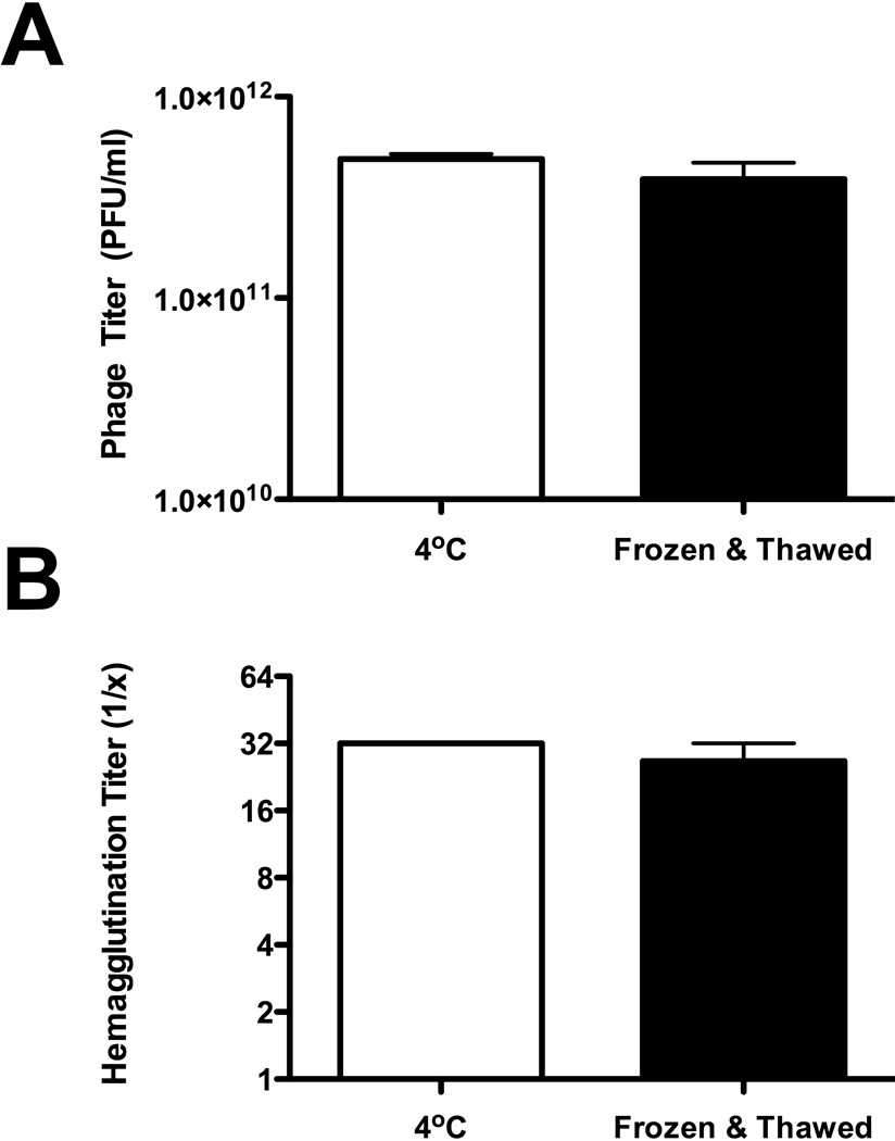
Finally, the physical stability of H5 HA-displaying phage particles was examined by subjecting them to a single freeze/thaw cycle, and then measuring the infectivity of the phage for bacterial target cells, as well as the ability of the phage particles to hemagglutinate turkey red blood cells (Figure 5). The results show that infectious phage titers and hemagglutination titers were unchanged by a single freeze/thaw cycle.
Figure 5. Phage capsids decorated with gpD:H5HA are stable after freeze/thaw.
H5 HA-decorated lambda phage particles were either stored overnight at 4°C or frozen at −80°C and then thawed. The infectivity of the phage for bacterial target cells was then measured (phage titration) (A), as well as the ability of the phage particles to hemagglutinate turkey red blood cells (B). Infectious phage titers (A) and hemagglutination titers (B) were unchanged by a single freeze/thaw cycle (Mann Whitney test, p = 0.4 [A] or 0.5 [B]).
At present, the overall turnaround time for producing an HA-displaying phage, starting from the cloning of a specified HA gene, is on order of 4–6 weeks. However, the longest step, by far, is the scaleup of baculovirus stocks for production of recombinant HA in insect cells. If one were to produce HA by transient transfection of mammalian cells, the overall turnaround time could be lowered to about a week – thereby greatly accelerating assay development. An additional potential advantage is the absence of NA on the phage particles. This may eliminate the need to conduct HAI assays at lower temperatures and/or allow assays to be remain stable for extended periods before being read.
In conclusion, these findings show that HA-displaying phage particles have utility as safe reagents for HAI assays on human sera in vaccine trials and other settings. This may have important advantages in the context of highly pathogenic viruses that require stringent biocontainment.
Supplementary Material
HIGHLIGHTS.
Bacteriophage lambda particles were coated with influenza virus H5 hemagglutinin (HA).
The HA coated phage particles agglutinated red blood cells efficiently.
A modified hemagglutination inhibition (HAI) assay was developed, using the particles.
This assay successfully identified human sera with H5 HA-specific antibodies.
HA-displaying bacteriophage capsids may be useful for detection of influenza-specific HAI antibodies.
Acknowledgements
This work was supported, in whole or in part, by National Institutes of Health (NIH) Contract HHSN266200700008C granted to the New York Influenza Center of Excellence. We thank Dr. Gary Nabel and the NIH Vaccine Research Center (VRC) for providing the 9E8 and 9B11 monoclonal antibodies, and we thank Dr. David J. Topham (University of Rochester) for valuable discussions. We also thank Theresa Fitzgerald for technical assistance.
Footnotes
Publisher's Disclaimer: This is a PDF file of an unedited manuscript that has been accepted for publication. As a service to our customers we are providing this early version of the manuscript. The manuscript will undergo copyediting, typesetting, and review of the resulting proof before it is published in its final citable form. Please note that during the production process errors may be discovered which could affect the content, and all legal disclaimers that apply to the journal pertain.
REFERENCES
- Baker SF, Guo H, Albrecht RA, Garcia-Sastre A, Topham DJ, Martinez-Sobrido L. Protection against lethal influenza with a viral mimic. J Virol. 2013;87:8591–8605. doi: 10.1128/JVI.01081-13. [DOI] [PMC free article] [PubMed] [Google Scholar]
- Gupta A, Onda M, Pastan I, Adhya S, Chaudhary VK. High-density functional display of proteins on bacteriophage lambda. J Mol Biol. 2003;334:241–254. doi: 10.1016/j.jmb.2003.09.033. [DOI] [PubMed] [Google Scholar]
- Hirst GK. The quantitative determination of influenza virus and antibodies by means of red cell agglutination. J Exp Med. 1942;75:49–64. doi: 10.1084/jem.75.1.49. [DOI] [PMC free article] [PubMed] [Google Scholar]
- Imperiale MJ, Hanna MG., 3rd Biosafety considerations of mammalian-transmissible H5N1 influenza. MBio. 2012;3:e00043–e00012. doi: 10.1128/mBio.00043-12. [DOI] [PMC free article] [PubMed] [Google Scholar]
- Katz JM, Hancock K, Xu X. Serologic assays for influenza surveillance, diagnosis and vaccine evaluation. Expert Rev Anti Infect Ther. 2011;9:669–683. doi: 10.1586/eri.11.51. [DOI] [PubMed] [Google Scholar]
- Keitel WA, Campbell JD, Treanor JJ, Walter EB, Patel SM, He F, Noah DL, Hill H. Safety and immunogenicity of an inactivated influenza A/H5N1 vaccine given with or without aluminum hydroxide to healthy adults: results of a phase I–II randomized clinical trial. J Infect Dis. 2008;198:1309–1316. doi: 10.1086/592172. [DOI] [PMC free article] [PubMed] [Google Scholar]
- Lipsitch M, Bloom BR. Rethinking biosafety in research on potential pandemic pathogens. MBio. 2012;3 doi: 10.1128/mBio.00360-12. [DOI] [PMC free article] [PubMed] [Google Scholar]
- Martinez-Sobrido L, Cadagan R, Steel J, Basler CF, Palese P, Moran TM, Garcia-Sastre A. Hemagglutinin-pseudotyped green fluorescent protein-expressing influenza viruses for the detection of influenza virus neutralizing antibodies. J Virol. 2010;84:2157–2163. doi: 10.1128/JVI.01433-09. [DOI] [PMC free article] [PubMed] [Google Scholar]
- Mattiacio J, Walter S, Brewer M, Domm W, Friedman AE, Dewhurst S. Dense display of HIV-1 envelope spikes on the lambda phage scaffold does not result in the generation of improved antibody responses to HIV-1 Env. Vaccine. 2011;29:2637–2647. doi: 10.1016/j.vaccine.2011.01.038. [DOI] [PMC free article] [PubMed] [Google Scholar]
- Mikawa YG, Maruyama IN, Brenner S. Surface display of proteins on bacteriophage lambda heads. J Mol Biol. 1996;262:21–30. doi: 10.1006/jmbi.1996.0495. [DOI] [PubMed] [Google Scholar]
- Santini C, Brennan D, Mennuni C, Hoess RH, Nicosia A, Cortese R, Luzzago A. Efficient display of an HCV cDNA expression library as C-terminal fusion to the capsid protein D of bacteriophage lambda. J Mol Biol. 1998;282:125–135. doi: 10.1006/jmbi.1998.1986. [DOI] [PubMed] [Google Scholar]
- Sternberg N, Hoess RH. Display of peptides and proteins on the surface of bacteriophage lambda. Proc Natl Acad Sci U S A. 1995;92:1609–1613. doi: 10.1073/pnas.92.5.1609. [DOI] [PMC free article] [PubMed] [Google Scholar]
- Treanor J. Recombinant proteins produced in insect cells. Curr Top Microbiol Immunol. 2009;333:211–225. doi: 10.1007/978-3-540-92165-3_11. [DOI] [PubMed] [Google Scholar]
- Treanor JJ, Campbell JD, Zangwill KM, Rowe T, Wolff M. Safety and immunogenicity of an inactivated subvirion influenza A (H5N1) vaccine. N Engl J Med. 2006;354:1343–1351. doi: 10.1056/NEJMoa055778. [DOI] [PubMed] [Google Scholar]
- Yang F, Forrer P, Dauter Z, Conway JF, Cheng N, Cerritelli ME, Steven AC, Pluckthun A, Wlodawer A. Novel fold and capsid-binding properties of the lambda-phage display platform protein gpD. Nat Struct Biol. 2000;7:230–237. doi: 10.1038/73347. [DOI] [PubMed] [Google Scholar]
- Yang ZY, Wei CJ, Kong WP, Wu L, Xu L, Smith DF, Nabel GJ. Immunization by avian H5 influenza hemagglutinin mutants with altered receptor binding specificity. Science. 2007;317:825–828. doi: 10.1126/science.1135165. [DOI] [PMC free article] [PubMed] [Google Scholar]
Associated Data
This section collects any data citations, data availability statements, or supplementary materials included in this article.


