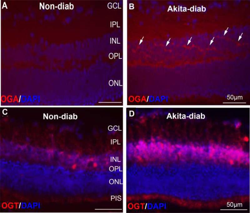
Figure 3.
OGA and OGT in diabetic Akita mouse retina. Retinal cryo-sections were prepared from the 5~6-month-old Akita diabetic mice or non-diabetic control mice. Immunofluorescence images of OGA in non-diabetic (A) and control (B) mice. Arrows indicate OGA-positive cells. Immunofluorescence images of OGT in non-diabetic (C) and control (D) mice. GCL: Ganglion cell layer; IPL: inner plexiform layer; INL: inner nuclear layer; OPL: outer plexiform layer; ONL: outer nuclear layer. Counterstaining with diamidino-2-phenylindole (DAPI) was used to highlight the three nuclear layers in the retina: ONL, INL, and GCL.

