
Figure 2.
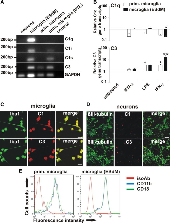
Complement production by microglia. A, RT-PCR of neurons, microglia (ESdMs) and primary (prim.) microglia. Transcripts for C1q, C1r, C1s, and C3 were detected in unstimulated microglia (ESdMs). C3 was constitutively transcribed in primary microglia, while C1r and C1s were detected after IFNγ stimulation. GAPDH served as standard. Control: PCR without cDNA. n = 3. B, Microglia (prim. and ESdMs) were untreated or stimulated for 24 h with IFNα (1000 U/ml), LPS (500 ng/ml), or IFNγ (100 U/ml). After LPS treatment transcripts of C3 were upregulated (prim. microglia: p = 0.015 vs untreated). After IFNγ stimulation transcripts of C1q were downregulated (ESdMs: p = 0.024 vs untreated), while C3 was upregulated (prim. microglia: p = 0.015 vs untreated; ESdMs: p = 0.001 vs untreated). n = 3 for prim. microglia (C1q), n = 5 for prim. microglia (C3), n = 4 for ESdMs. C, D, Microglia and neurons were double-immunostained with antibodies directed against C1q/C3 and Iba-1/βIII-tubulin. Protein expression of C1q and C3 was detected in microglia while it was undetectable in neurons. Scale bar, 20 μm, n = 3. E, Flow cytometry of microglia (prim. and ESdMs). Expression of CD11b (blue) and CD18 (green) was detected at similar levels by prim. microglia and microglia (ESdMs). An isotype antibody (red) was used as negative control. n = 3.