
Figure 4.
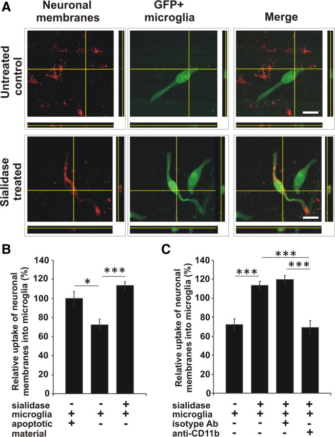
Uptake of desialylated neurons by microglia. Microglia transduced with GFP (GFP+ microglia) were cocultured with normal (untreated control) or sialidase-treated neurons, which were labeled with a red fluorescent membrane dye. Uptake of neuronal membranes into microglia was visualized by confocal microscopy and 3D-reconstruction. Uptake of apoptotic red fluorescent labeled material added to the coculture system served as positive control. A, After coculture of microglia with desialylated neurons fluorescent-marked neuronal membranes were detected inside the microglia. Scale bars, 20 μm, n = 3. B, C, Quantification of uptake. Few neuronal membranes were detected in microglia cocultured with normal neurons (untreated). Uptake of red fluorescent neuronal membranes into microglia was increased (***p = 4.8 × 10⁻⁵) after desialylation of the neuronal glycocalyx (sialidase treated). Few uptake of neuronal membranes was observed after blocking CD11b (***p = 7.27 × 10⁻⁶ vs +sialidase, ***p = 1.9 × 10⁻⁷ vs +sialidase+isotype Ab). n = 3.