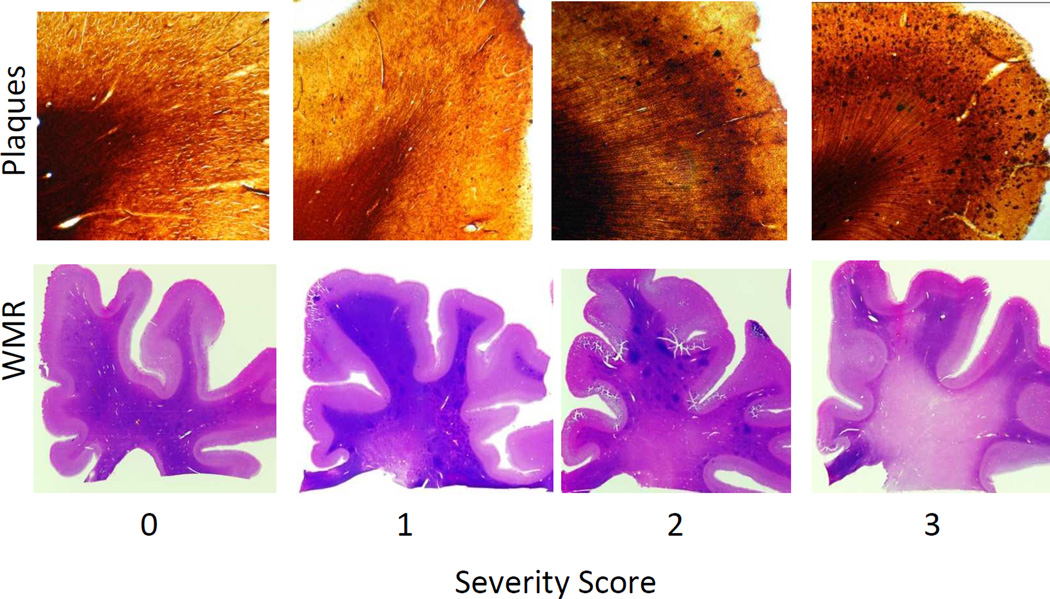
Figure 1.
Examples of severity scores given to plaque densities (top) and white matter rarefaction (WMR- bottom). Top: 80 µm sections of the superior frontal gyrus stained with the Campbell Switzer enhanced silver stain -from left to right: 0 - none, 1 - mild, 2 - moderate, and 3 - frequent plaque densities. Bottom: macro view of 80 µm sections of the frontal lobe stained with hematoxylin and eosin, WMR was scored as from left to right: 0 - none, 1 - mild, 2 - moderate, and 3 - severe. In this study, a case was defined as having significant WMR if it had a score of 2 or higher in one or more of the following lobes: frontal, parietal, temporal and occipital.

