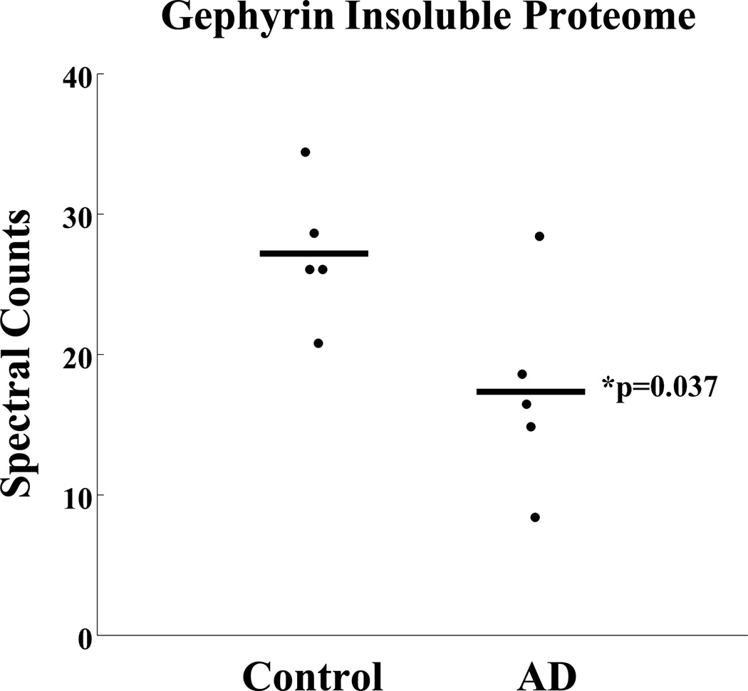
Figure 6. Full-length gephyrin expression is reduced in AD insoluble proteome.
The insoluble fraction (urea soluble) from 5 control and 5 AD cases (including cases in Figure 5) were subject to mass spectrometry analysis. Samples were resolved on an acrylamide gel and lanes were divided into 5 different molecular weight regions. Gephyrin peptides were identified in the molecular region consistent with full-length gephyrin. Spectral counts (number of gephyrin peptides sequenced) were normalized following removal of system background. The AD mean is significantly reduced compared to controls (p-value <0.05).

