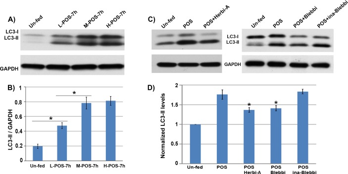
Figure 7.
(A) Western blot of lysates from RPE-J cells exposed to various amounts of POS. L-, M-, and H- correspond to 2, 10, or 50 outer segments per RPE cell, respectively. (B) Quantification of the LC3-II levels based on the densitometry of the Western blots. The level of LC3-II appears to peak with the medium dose of 10 outer segments per RPE cell (P < 0.05). (C) Western blot of lysates from RPE-J cells fed POS with or without the addition of the phagocytosis inhibitors herbimycin-A (Herbi-A) or blebbistatin (Blebbi). As a control, an inactive form of blebbistatin (Ina-Blebbi) was used, and found to have no effect on LC3-II formation. (D) Quantification of the LC3-II levels based on the densitometry of the Western blots. The levels of LC3-II in the herbimycin-A and blebbistatin treated cells were significantly lower than control-treated cells (P < 0.05).

