Figure 5.

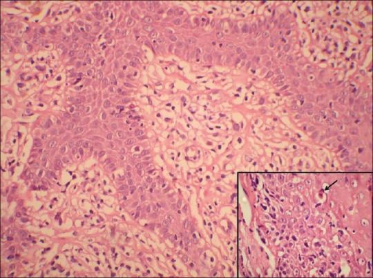
Photomicrograph showing basal cell vacuolization and lymphocytic infiltrate at dermo-epidermal junction. (H and E, ×400). Inset showing civatte body

Photomicrograph showing basal cell vacuolization and lymphocytic infiltrate at dermo-epidermal junction. (H and E, ×400). Inset showing civatte body