Figure 6.

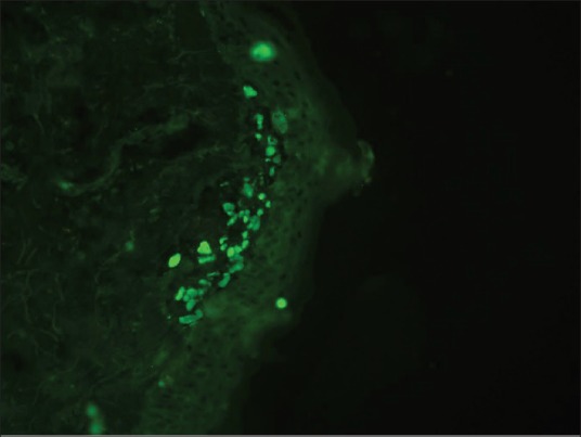
DIF photomicrograph showing IgM reactive large grouped globular (++) deposits (Civatte body) in the papillary dermis (anti-IgM, ×400)

DIF photomicrograph showing IgM reactive large grouped globular (++) deposits (Civatte body) in the papillary dermis (anti-IgM, ×400)