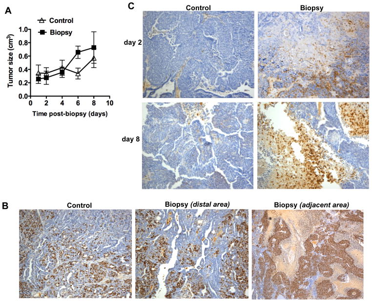
Figure 1. Effect of mammary tumor biopsy in tumor progression.
(A) Size of the tumors that underwent biopsy (Biopsy) and control tumors (Control) over time after biopsy (n=6 mice). Tumor growth rate was slightly increased in mice undergoing biopsy (p < 0.05), as determined by random coefficient analysis with time included as a quadratic term. (B) Immunohistochemistry staining for Ki67 in tumors isolated from “Control mice” (control) and “Biopsy mice” (Biopsy) 8 days after biopsy. For “Biopsy mice”, an area distal to the biopsy and an area adjacent to the biopsy site within the same tumor are shown. 100x magnification. (C) Immunohistochemistry staining for CD45 in sections of tumors isolated from Control mice and Biopsy mice that had biopsy 2 days or 8 days prior to tissue harvesting. 100 × magnification.

