
Figure 1.
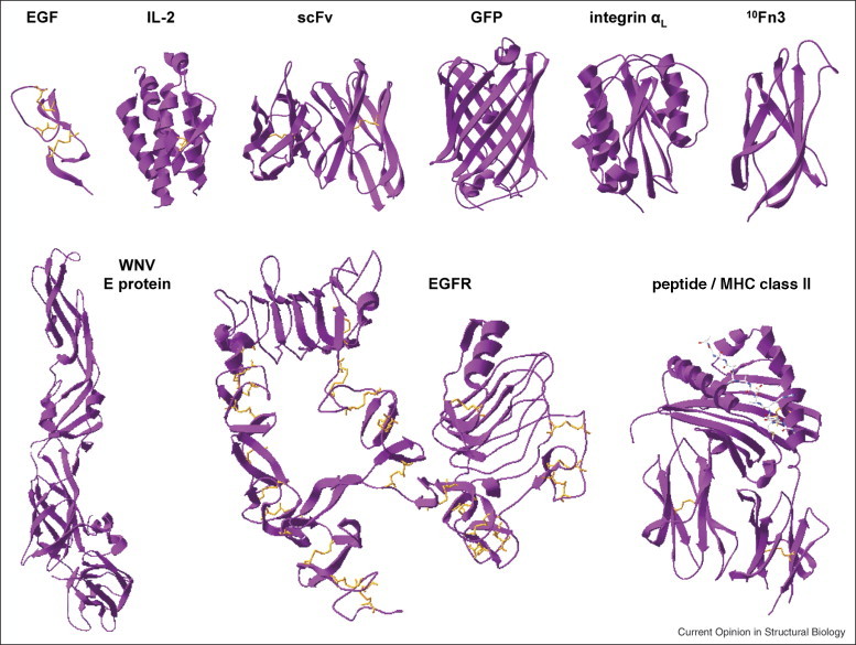
A selection of proteins successfully displayed as Aga2p fusions on the surface of yeast. Top row, left to right: human epidermal growth factor [31••] (1JL9), human interleukin-2 [61] (2B51), single-chain antibody fragment 4m5.3 [62] (1X9Q), green fluorescent protein [63] (1EMA), human αL integrin inserted domain [19] (1LFA), and human fibronectin [18] (1FNA). Bottom row, left to right: West Nile Virus envelope protein [32•] (2I69), human EGF receptor ectodomain [27] (1NQL), and human MHC class II HLA-DR4αβ in complex with peptide [64] (2SEB). The PDB IDs for the structures shown are noted in parentheses. This figure was generated using Swiss-Pdb Viewer [65].