Figure 5.

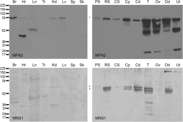
Immunoblot of somatic and reproductive tissues probed with antibodies against MFN2 (top) or MNS1 (bottom). MFN2 was seen in brain, heart, kidney, and liver (top, left panel) and was found in all reproductive tissues examined (top, right panel). MNS1 was seen faintly in the kidney (bottom, left panel) and was present as two closely migrating bands in all reproductive tissues and isolated cells examined except pachytene spermatocytes, condensing spermatids, and uterus (bottom, right panel). Br: brain, Hr: heart, Ln: lung, Tr: trachea, Kd: kidney, Lv: liver, Sp: spleen, Sk: skeletal muscle, PS: pachytene spermatocytes, RS: round spermatids, CS: condensing spermatids, Cp: caput epididymal sperm, Cd: epididymal sperm, T: testis, Ov: ovary, Od: oviduct, Ut: uterus. Numbers to the left of the panels indicate the molecular weights of standard proteins (×10-3). Asterisk (*) indicates expected size (MFN2: M r of 80 and MNS1: M r of 60) of protein.
