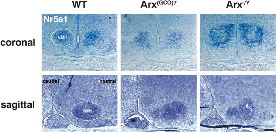
Figure 1. Nr5a1 demarcates the VMH in WT and Arx transgenic mice.

Representative coronal and sagittal sections, probed with Nr5a1, are shown for wild type (left panels), Arx(GCG)7/Y (middle panels) and Arx−/Y (right panels) mice. For reference, the left ventromedial hypothalamic nucleus (VMH) is roughly enclosed by dotted lines in the wild type images, but is present in all sections probed. Scale bar = 200 µm.
