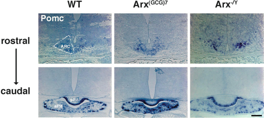
Figure 2. Pomc demarcates the ARC and pituitary in WT and Arx transgenic mice.

Representative coronal sections, probed with Pomc, are shown for wild type (left panels), Arx(GCG)7/Y (middle panels) and Arx−/Y (right panels) mice. In the wild type images, the approximate outline of the left arcuate nucleus (ARC) is illustrated by dotted lines in the top, more rostral section, whereas the anterior and posterior pituitary (aPit and pPit, respectively) are labeled in the bottom, more caudal section. These nuclei are also normally expressing Pomc in the Arx transgenic animals. Scale bar = 200 µm.
