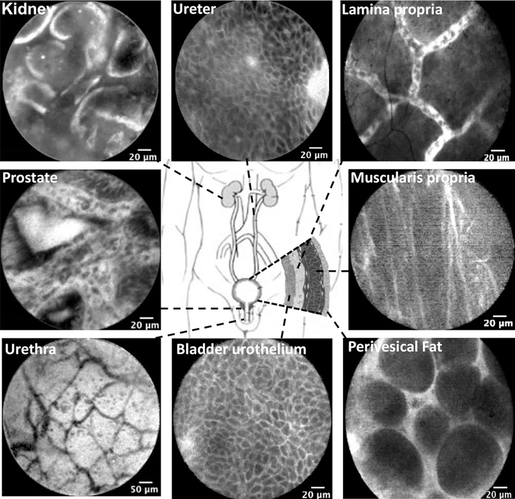
Figure 2.
Probe-based confocal laser endomicroscopy (pCLE) images of the normal urinary tract. All images of the lower urinary tract were acquired in vivo, whereas upper tract images (kidney cortex and ureter) were acquired ex vivo. Note similarities of the urothelium (intermediate cells) between ureter and bladder. Lamina propria is characterized by capillary network of moving erythrocytes. Muscularis propia and perivesical fat images were obtained from the tumor resection bed.

