Figure 5.

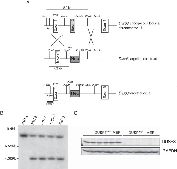
Dusp3 deficient mice generation by targeted homologous recombination. (A) Schematic diagram showing part of the Dusp3 gene locus, the targeted Dusp3 construct and the resulting targeted allele. Recombination events are indicated by open white boxes and show the replacement of a 8.2 kb Dusp3 genomic fragment containing exon II by the pPNT-Neo cassette. (B) Southern blot analysis of ES cells genomic DNA following digestion with XbaI using a 5’ external Probe P as indicated in (A). The autoradiography revealed the 8.2 kb (wild-type) and 4.5 kb (targeted) fragments. The stars represent the ES cell lines used for microinjection of mouse blastocysts. (C) Western blot analysis of DUSP3 protein expression in MEF cell extracts from 6 Dusp3+/+ and 6 Dusp3−/− mice. GAPDH was used as an internal control.
