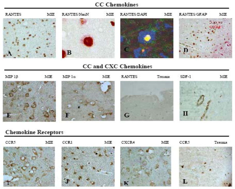
Figure 1. Expression of CC and CXC chemokines and chemokine receptors in MIE (medically intractable temporal lobe epilepsy) and trauma.

Immunohistochemistry or confocal microscopy of temporal lobe (TL) and hippocampus (H) using the following antibodies: (A) RANTES (TL, 40X); (B) RANTES/NeuN (TL, 100x); (C) RANTES green /DAPI (TL, confocal microscopy 100x) (D) RANTES/GFAP (H, 40x); (E) MIP -1 β (40x); (F) MIP-1α(40x); (G) RANTES (Trauma cortex, 20x); (H) SDF-1(H,40x); (I) CCR5 (20x); (J) CCR1 (40x); (K) CXCR4 (40x); (L) CCR5 (Trauma cortex, 40x). Note RANTES expression in neuronal nuclei (A-D) but cytoplasmic expression of MIP-1α and MIP-1β extending into the neuropil (E, F).
