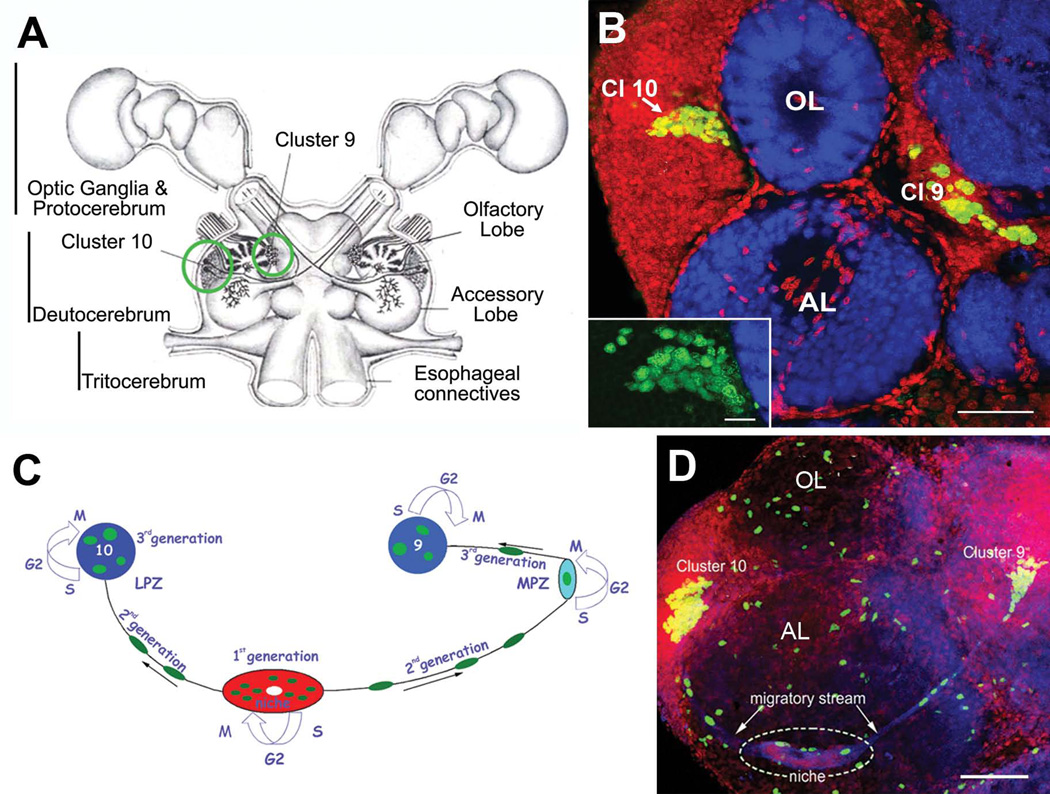
Figure 1.
Neurogenesis in the adult crayfish (C. destructor) brain. (A) Schematic diagram of the crayfish brain. Cell clusters 9 and 10 (circled in green), where adult-born neurons are incorporated, flank the olfactory and accessory lobes of the deutocerebrum. (B) Horizontal section through the olfactory (OL) and accessory lobes (AL) of C. destructor labeled immunocytochemically for BrdU (green) and Drosophila synapsin (blue) and counterstained with propidium iodide (red), a marker of nucleic acids. BrdU-labeled cells are observed within the proliferation zone in soma cluster 10 (Cl 10) (arrow), which lies adjacent to the olfactory lobe and in cluster 9 (Cl 9). The inset shows a higher-magnification view of BrdU-labeled cells within the cluster 10 proliferation zone. (C) A model summarizing our current understanding of events leading to the production of olfactory interneurons in adult crayfish. First generation neuronal precursor cells reside in a neurogenic niche where they divide symmetrically. Their daughters (second-generation precursors) migrate towards the lateral proliferation zone in Cluster 10 (LPZ) or the medial proliferation zone (MPZ) in Cluster 9 along tracts created by the fibers of bipolar niche cells. At least one more division occurs in the LPZ and MPZ before the progeny (third and subsequent generations of precursors) differentiate into neurons. (D) Left side of the brain of Procambarus clarkii labeled immunocytochemically for the S-phase marker BrdU (green). Labeled cells are found in the lateral proliferation zone contiguous with Cluster 10 and in the medial proliferation zone near Cluster 9. The two zones are linked by a chain of cells in the migratory stream, which labels immunocytochemically for glutamine synthetase (GS; blue). These streams originate in the oval region ‘niche’ (dotted circle) containing cells labeled with the nuclear marker propidium iodide (PI, red). The BrdU-labeled cells scattered irregularly throughout the OL and AL (which do not contain neuronal cell bodies) are glial cells. Scale bars: 100 µm in (B); 20 µm in insert in (B); 75µm in (D).

