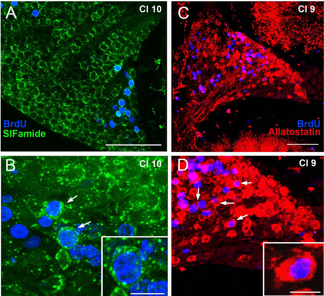
Figure 4.
Differentiation of cluster 9 and cluster 10 BrdU-labeled cells. (A, B) Immunocyto-chemical labeling of cluster 10 cells for BrdU (blue) and SIFamide (green). By week 8, many BrdU-labeled cells are co-labeled with SIFamide (arrows in B), indicating that these have differentiated into neurons. (C, D) Images of BrdU (blue) and allatostatin (red) labeling of cluster 9 neurons (arrows), 8 weeks after the BrdU-labeling period. Scale bars = 100µm in A and B; 15µm in insert in B and D.

