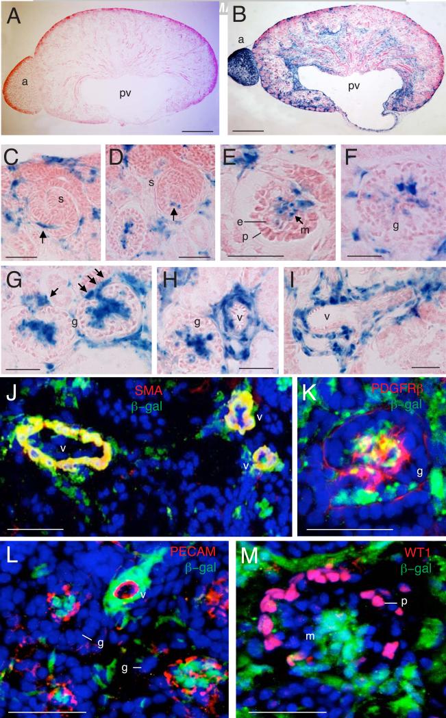
Fig. 2. Contribution of Tbx18-derived progenitors to renal vascular system and interstitium.
(A,B) X-gal staining on kidney sections of R26RLacZ (A) or Tbx18Cre;R26RlacZ (B) embryos at E17.5. (C,D) Higher magnification of X-gal stained sections from Tbx18Cre;R26RlacZ embryos at E17.5 showing Tbx18-derived cells are associated with the capillary sprout entering into the S-shaped glomerulus (s) (arrows). (E) Early, (F) mature and (G) late cup-shaped glomerular mesangial cells are derived from Tbx18+ cells but podocytes and endothelial cells are not linearly related to Tbx18+ cells. (H,I) Perivascular mesenchyme is originated from Tbx18+ cells. (J) Co-immunostaining with β-gal and α-SMA antibodies showing that vascular α-SMA+ cells are vascular β-gal+ (arrows). (K) Co-immunostaining with β-gal and PDGFRβ antibodies showing glomerular mesangial cells double positive for E -gal and PDGFRβ (arrows). (L,M) Coimmunostaining with β-gal along with PECAM-1 and WT1 antibodies showing no E -gal+ cells are positive these two markers glomeruli (g) and vessels. Other abb.: g, m, mesangial cells; v, vessels. Scale bar: 200 μm for A,B, 50 μm for C-M.

