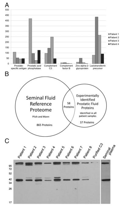
FIGURE 1.
Proteomic analysis of prostatic fluid samples from radical prostatectomy specimens of men with prostate cancer. (A) Examples of the 95 proteins identified in each of four prostatic fluid samples. Y-axis indicates number of peptide “hits” for each protein from mass spectrometric analysis. (B) The prostatic fluid experimental dataset has considerable overlap with the previously described seminal plasma reference database. A Venn diagram was constructed showing overlap between protein species identified in our screen of prostatic fluid samples and the seminal plasma reference proteome of Pilch and Mann (12). Protein species found in our screen must have been identified in all four prostatic fluid samples to be included in the comparison. (C) Patient prostatic fluid and normal seminal plasma contains native C3 and a C3 fragment at ~37 kDa. Western blot of eight random prostatic fluid samples, purified human C3, and the seminal plasma of a healthy donor were probed for the presence of C3 with a monoclonal anti-human–C3b-α (clone H206) Ab.

