Fig. 5.
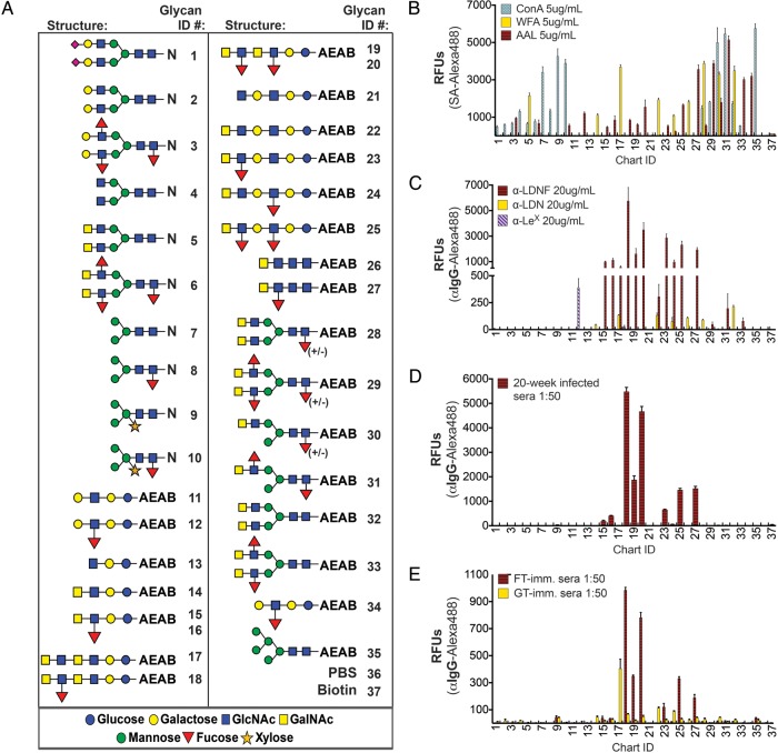
Antibodies to schistosome glycans discriminate among very similar presentations of the epitope. (A) N-linked glycopeptides and AEAB-linked glycans were modified to include several variants of LDN, LDNF and other schistosome antigens and printed on glass slides, called the defined schistosome-type microarray (DSA). LDN-terminating glycans are printed at 50 μM and all other glycans are printed at 100 μM due to variation in reaction yields. Some LDNF-terminating glycans are printed at 50 and 100 μM, in that order, for comparison with LDN. (B) Biotinylated lectins binding tri-mannose (ConA), terminal GalNAc (WFA) and fucose (AAL) were used to quality-control printing of the slides. (C) Anti-schistosomal monoclonal antibodies, (D) antisera from chronically S. mansoni-infected mice and (E) week 20 antisera from L8-GT and L8-GTFT (FT) immunization were tested on the DSA. Streptavidin-Alexa488 or goat anti-mouse-IgG-Alexa488 were used to detect biotinylated lectins and antibodies bound to the slides, respectively. Mean RFUs ± SD of tetra-replicate spots for each glycan ID are shown. N, asparagine; AEAB, 2-amino-N-(2-aminoethyl)-benzamide; RFUs, relative fluorescence units; SA, streptavidin; ConA, concanavalin A; WFA, Wisteria floribunda agglutinin; AAL, Aleuria aurantia lectin.

