Fig. 6.
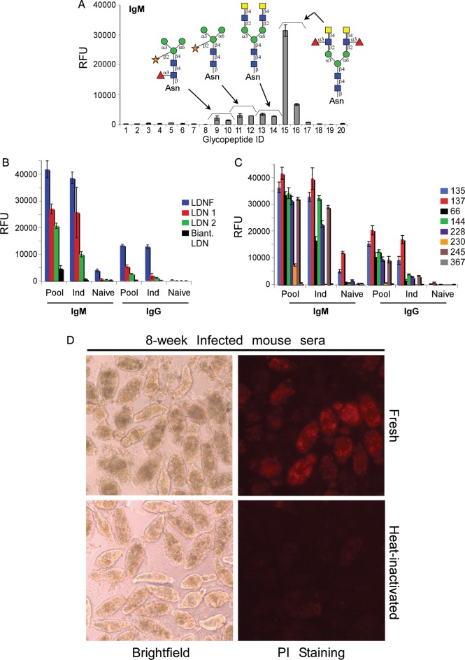
Antibody responses and complement-dependent killing activity in infected mice. (A) Sera from 8 week-infected mice were used to probe the defined schistosome glycan microarray for IgM binding (IgG data not shown). (B and C) Sera from infected mice were used to probe a CFG glycan microarray. IgM and IgG (B) responses to LDNF and various LDN structures, and IgM and IgG (C) responses to various LeX epitopes are shown from the following samples: pooled (Pool), individual (Ind) and an individual naïve mouse. Numbers in the legend for C correspond to the glycans listed in Table II. (D) Mechanically transformed schistosomula were treated with pooled sera from 8-week S. mansoni infected mice diluted in culture medium to 1:10. Death of schistosomula was assessed by granularity and loss of membrane integrity (brightfield) and fluorescent propidium iodide (PI) uptake. Data are representative of two replicate experiments. Serum was either fresh (obtained from mice the same day and kept at room temperature) or heat-inactivated to destroy complement at 56°C for 1 h. A section of an image taken at 10× is enlarged to show localization of PI stain to individual dead schistosomula.

