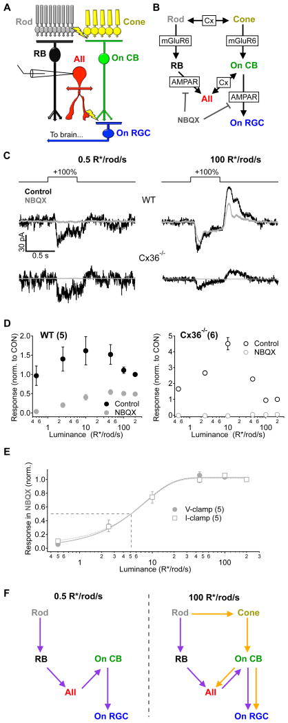
Figure 3. Signal propagation through parallel circuits depends on luminance.
A, Simplified diagram of the parallel circuits that transmit visual information to On alpha RGCs in mouse retina. B, Detailed signaling schematic of the circuit elements and synaptic receptors that mediate transmission within the On alpha RGC circuit. Cx - gap junctions containing connexin proteins, mGluR6 - metabotropic glutamate receptor 6, AMPAR - fast ionotropic glutamate receptors. C, Responses to +100% contrast steps measured in voltage clamp in an AII amacrine cell from both WT (top) and Cx36−/− (bottom) mice before and after bath application of NBQX (10 μM). D, AII amacrine cell responses across luminance (± NBQX) normalized to the control response at 200 R*/rod/s in wild type (WT, left) and Cx36−/−(right) retinas. Responses from AII amacrine cells in Cx36−/− mice were completely eliminated in the presence of NBQX, confirming a decoupling of the two pathways. E, Responses recorded from AII amacrine cells in the presence of NBQX in the voltage clamp configuration (same data is in D; closed circles) or the current clamp configuration (open squares) normalized to the response at 200 R*/rod/s. Fits are sigmoids with the half-max indicated by dotted line. The background eliciting a half-maximal response to 100% contrast steps in the presence of NBQX was 5.1 R*/rod/s, regardless of the recording configuration. F, Schematic illustrating the flow of signals through the rod bipolar (purple) and cone bipolar (orange) circuits at two different luminance levels. Error bars in D and E are s.e.m. across cells. Numbers of cells are indicated in parentheses in panel legends. All recordings are from slice preparations.

