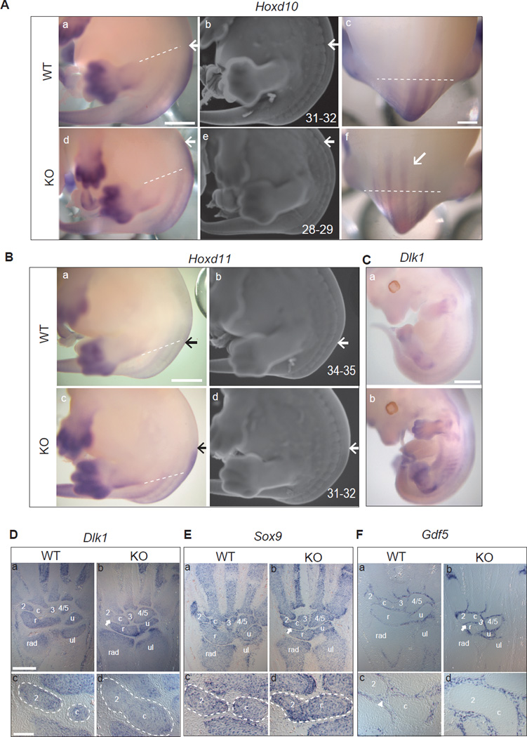
Figure 3. Spatial and temporal gene expression patterns in Hotair KO mice.
(A-B) Whole mount in situ hybridization (WISH) of Hoxd10 (A-a/c/d/f) and Hoxd11 (Ba/ c) of E13.5 embryos (n>3 for each genotype). KO embryos showed increased intensity and anterior shift of the expression domains of HoxD genes (highlight with arrows, the dotted lines across hind limbs are used as anatomical limit). Same embryos were co-stained with ethidium bromide, and the somite position of the anterior expression domain were numbered and marked with arrows (A-b/e; B-b/d). Scale bar: 1mm for A-a/b/d/e, B-a/b/c/d; 600µm for A-c/f.
(C) WISH of Dlk1 on E12.5 embryos showing ectopic expression in Hotair KO embryos. (WT, n=4; KO, n=5; scale bar: 1mm).
(D, E, F) Altered Dlk1 expression and mesenchymal cell fates in Hotair KO wrists. Dlk1 (D), Sox9 (E) and Gdf5 (F) expression in E15.5 wrist sections (n>3 for each genotype). Arrows indicate the joint regions in KO. Dotted circles marked the carpal element 2 and central element c. Arrowhead indicates the intervening Gdf5-positive domain in WT. Note 2-c fusion in KO wrist, showing continuous Dlk1 and Sox9 expression in the junction area of 2 and c; and loss of Gdf5 signal as well. (Scale bar: 300µm for D-a/b, E-a/b, F-a/b; 100µm for D-c/d, E-c/d, F-c/d)
See also Figure S3.

