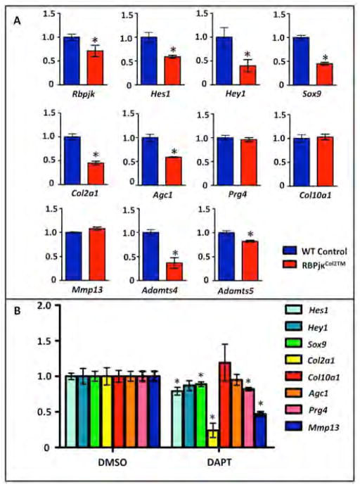
Figure 6. Impaired Notch signaling within articular chondrocytes results in rapid chondrocyte gene expression changes.
(A) Real-time qPCR comparing gene expression in the articular chondrocytes isolated from P30 WT and RBPjκCol2TM mutants following 5 days of TM administration (P25–29) and (B) WT cultured articular chondrocytes following two days of DAPT or DMSO treatment. Gene expression analyses were performed for Rbpjk, Hes1, Hey1, Sox9, Col2a1, Agc1, Prg4, Col10a1, Mmp13, Adamts4 and Adamts5. Bars represent means +/− SD (n=3). All samples are normalized to Beta-actin and then normalized to the controls. “*” denotes statistical significance with p-value less than 0.05. All assays were performed in triplicate.

