
Figure 3.
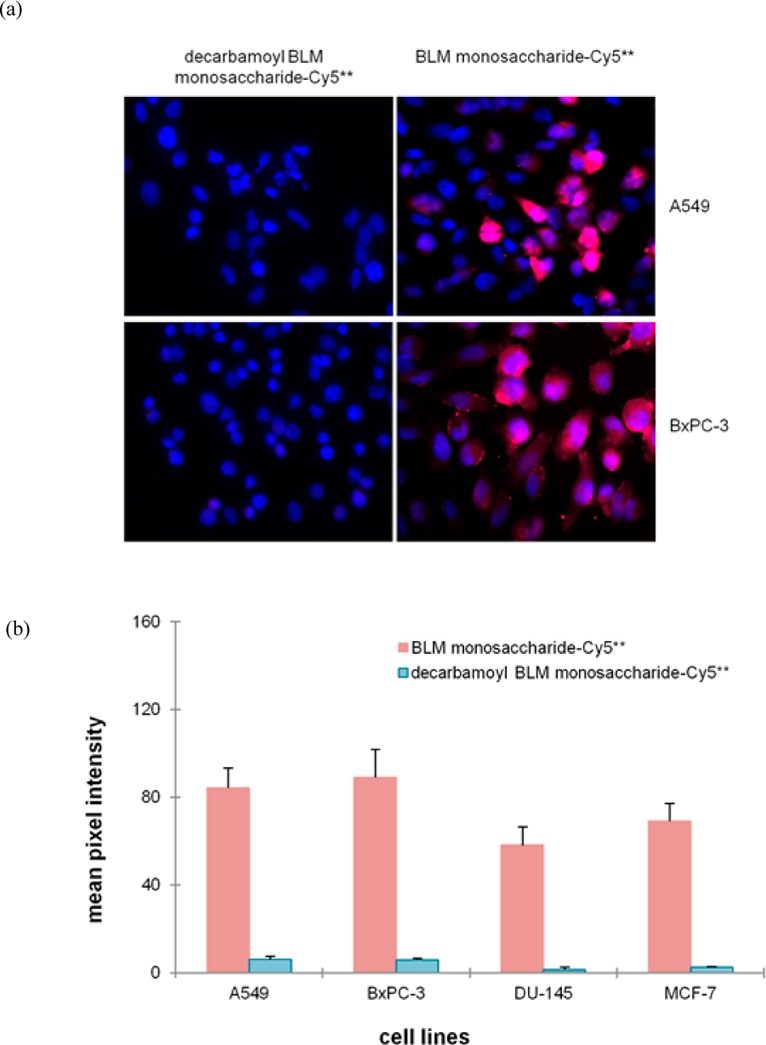
(a) Binding/uptake of decarbamoyl BLM monosaccharide–Cy5** and BLM monosaccharide–Cy5** conjugates in A549 and BxPC-3 cell lines. The cells were treated with 25 μM decarbamoyl BLM monosaccharide–Cy5** or BLM monosaccharide–Cy5** conjugate at 37 °C for 1 h, washed and fixed. The nuclei were stained with DAPI. Fluorescence imaging was conducted with a 3 s exposure time. (b) Quantification of the binding/uptake of decarbamoyl monosaccharide–Cy5** and BLM monosaccharide–Cy5** conjugates in four cancer cell lines. The cells were treated with 25 μM dye conjugates and irradiated for 3 s prior to being imaged.