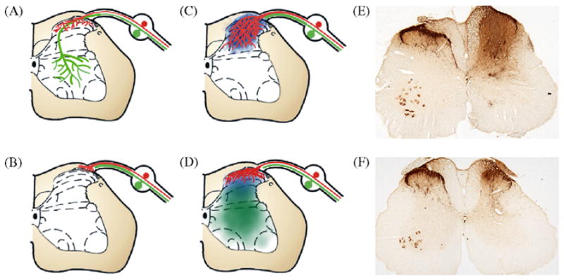
Fig. 1.

Recapitulation of developmental-like guidance cues can target regenerating sensory axons. Schematic illustration of the DREZ model used to examine regeneration of sensory afferents into the spinal cord. (A) Peptidergic nociceptive axons (red) in the normal spinal cord terminate in laminae I and II of the dorsal horn; whereas, large myelinated axons (green) extend to more ventral regions. (B) Dorsal root rhizotomy results in severing of both nociceptive and proprioceptive axons. These axons are capable of regenerating in the peripheral nerve up to, but not into, the spinal cord. (C) Injecting NGF adenovirus into the dorsal horn of the spinal cord (blue) supports the regeneration of sensory afferents through the DREZ and throughout the entire dorsal horn of the spinal cord, a pattern unlike normal. (D) To target regenerating axons to their normal laminae, NGF adenovirus was injected dorsally, while semaphorin 3A-encoding adenovirus was injected ventrally. This produced slightly overlapping gradients that supported regeneration of peptidergic nociceptive axons into the upper dorsal horn exclusively, generating a reinnervation pattern very similar to normal. (E) Photo of spinal cord treated with NGF dorsally and GFP ventrally (right side, as in C) showing extensive regeneration of sensory nociceptive axons throughout the entire dorsal horn. (F) Photo of spinal cord treated with NGF dorsally and Semaphorin 3A ventrally (right side, as in E) axon regeneration is restricted to the upper dorsal horn similar to the sham lesion side (left side of cord in E and F; Tang et al. [74]).
