Figure 5.

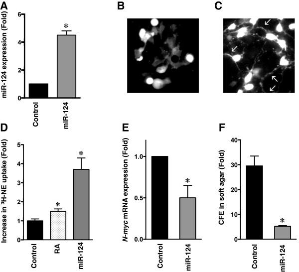
miR-124 induces neuronal differentiation in I-type NB cells. A. The miR-124 levels in control and miR-124-infected BE(2)-C. B. Immunofluorescence microscopy of BE(2)-C cells infected with a lentiviral-vector expressing GFP (control) or (C) BE(2)-C cells infected with lentiviral vector co-expressing miR-124 and GFP. Photomicrographs were taken two weeks after infection. Note the increase in number and size of multiple neuritic processes in C (arrows). D. 3H-NE uptake in BE(2)-C cells is increased 1.5–fold (P < 0.002) following treatment with RA and 3.7-fold (P < 0.001) with miR-124 lentiviral infection. E. N-myc mRNA levels are decreased ~2-fold (P < 0.008) in BE(2)-C/miR-124 lentiviral vector cells compared to control. F. Colony forming efficiencies (CFE) of BE(2)-C cells stably infected with miR-124 lentiviral vector or control. Note that CFE is reduced nearly 6-fold following infection (P < 0.001).
