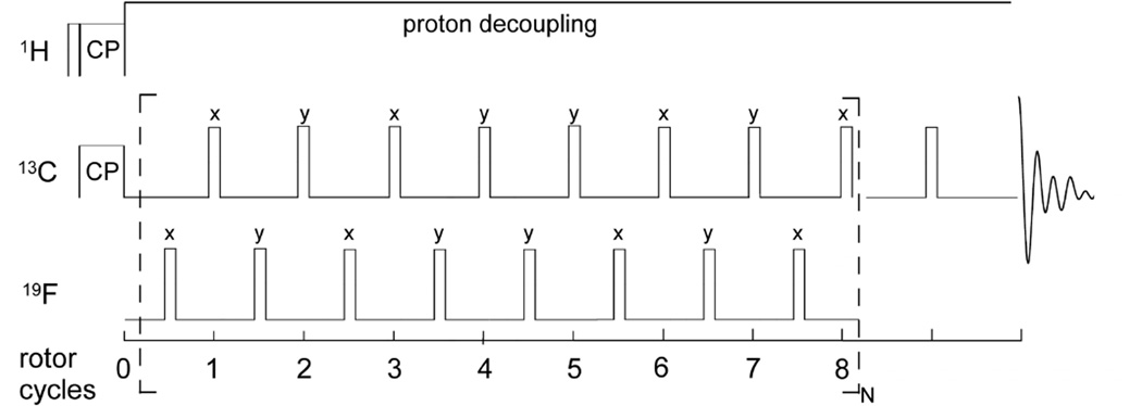
Figure 1. The REDOR measurement.
REDOR is performed in two parts, once with dephasing pulses (S spectrum) and once without (full echo, S0 spectrum). REDOR spectra are typically collected with standard xy-8 phase cycling55, on both observed and dephasing channels.

