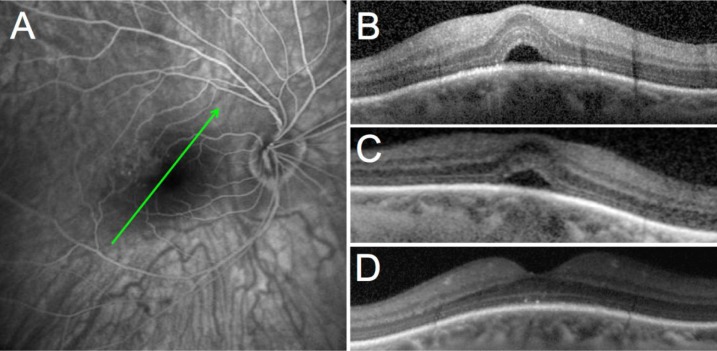
Figure 1.
Case 1: 53-year-old female patient.
Notes: (A) Late phase fluorescein angiography showing no sign of choroidal neovascularization or other causes of vascular leakage; (B) macular OCT of the right eye at presentation; (C) macular OCT after treatment with acetazolamide; (D) macular OCT after treatment with spironolactone. The green arrows indicate the level of the B-scan OCT sections.
Abbreviation: OCT, optical coherence tomography.

