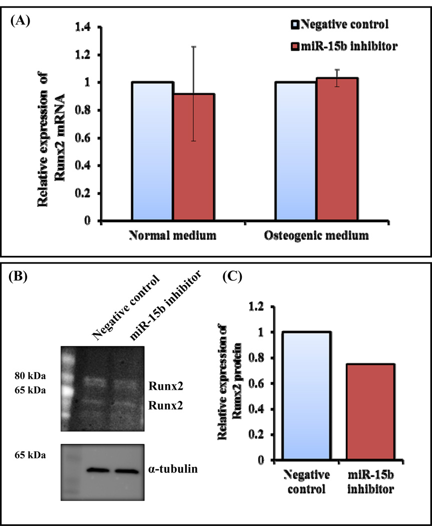
Figure 3.
Regulation of Runx2 by miR-15b inhibitor during hMSCs differentiation towards osteoblasts. hMSCs were transiently transfected with 50 nM of negative control miRNA or miR-15b inhibitor in the presence of normal medium or osteogenic medium. (A) At day 10, total RNA was isolated and real time RT-PCR was carried out using the primers for Runx2 and GAPDH genes. Relative expression of Runx2 mRNA was calculated after normalization with GAPDH. (B) At day 10, whole cell lysates were prepared and subjected to Western blot analysis using the antibody for Runx2. α-tubulin was used as internal control. (C) The densitometry scanning of the above Western blot after normalization with α-tubulin. The experiment was carried out three times and a representative blot is given.

