
Figure 8. Ras transformation affects the circadian clock via MAPK signalling.
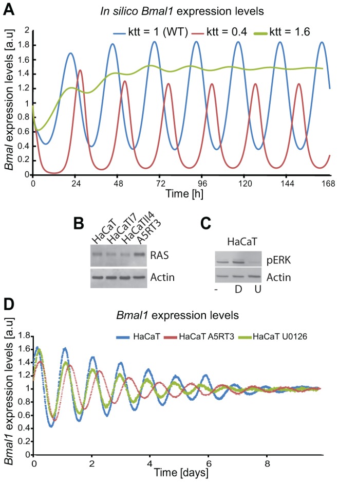
(A) Shown are in silico-generated gene expression oscillations with wild type (WT) in blue, RAS overexpressing in red (i.e., reducing the BMAL1 mediated-transcription by 60% (ktt = 0.4) compared to WT) and reducing RAS activity (i.e., increasing BMAL1 mediated-transcription by 60% (ktt = 1.6)) in green. (B) Western Blot analysis of RAS protein expression for the HaCaT cell lines and its derivatives (HaCaTI7, HaCaTII4, A5RT3), shows clear RAS overexpression measured in A5RT3. (C) Phosphorylated ERK (pERK) is shown for HaCaT cells under different conditions: non-treated cells, (−), cells treated with DMSO (D), cells treated with U0126 (U). HaCaT cells were treated with a MEK inhibitor (UO126 20 µmol) after synchronization with dexamethasone and circadian activity of Bmal1-luciferase reporter was measured over 8 days. (D) Shown are the results from 3 independent experiments. Non-treated (NT) HaCaT have a period of 22.9±0.2 hours (p<0.05, Student's t-test), HaCaT A5RT3 present a larger period of 24.5±0.1 hours (p<0.05, Student's t-test). Cells treated with UO126 (20 µM) show a period decrease (22.2±0.1 hours, p<0.05, Student's t-test) and a phase advance.