FIGURE 1:

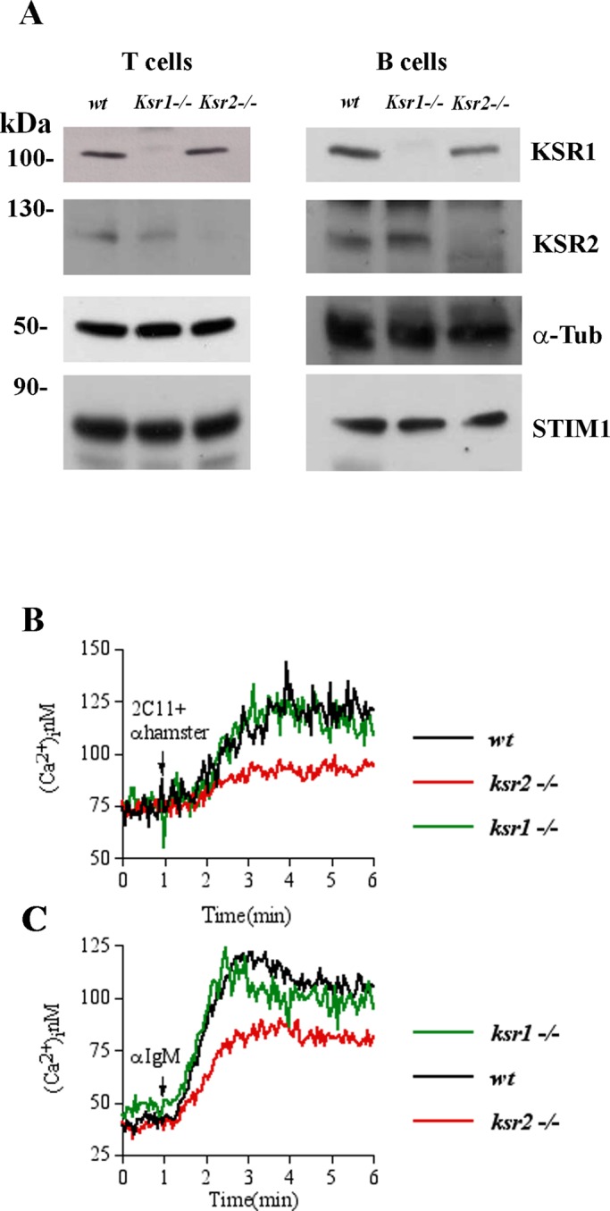
Defective [Ca2+]i elevation in ksr2−/− lymphocytes. (A) Immunoblot analysis of KSR1, KSR2, and STIM1 protein expression in purified T- and B-lymphocytes (15 × 106 cells/condition) from wt, ksr1−/−, and ksr2−/− mice. α-Tubulin (α-Tub) was used as loading control. Left, relative molecular mass (kilodaltons). (B) Elevation of [Ca2+]i induced by anti-CD3 (2C11) followed by anti-IgG antibody (anti-hamster) stimulation was assessed by spectrofluorimetric analysis with Fura-2 in 3 × 10⁶ T-cells purified from wt (black line), ksr1−/− (green line), and ksr2−/− mice (red line). Cells were kept in 1.0 mM Ca2+-containing medium. (C) Elevation of [Ca2+]i induced by anti-IgM stimulation performed in 3 × 10⁶ purified B-cells, as described in B. Traces represent one of three independent experiments.
