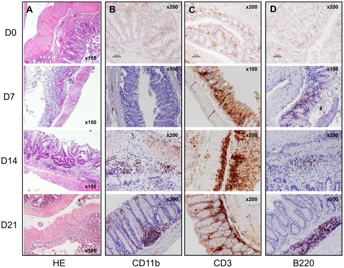
Figure 4. Histological and immunohistochemistry analyses of the Ncf1* colon show ulcers, epithelial hyperplasia and severe dysplasia, with massive infiltration of CD3+ lymphocytes, and subsequently of B220+ B lymphocytes and Mac-1+ cells in the later time-points.
A) Hematoxylin-eosin (HE) staining of the colon during the different phases of the DSS-induced colitis protocol. B) Immunohistochemistry of Mac-1+ cells in the inflammatory infiltrates at days 0, 7, 14 and 21. C) Immunohistochemistry of CD3+ cells in the inflammatory infiltrates at days 0, 7, 14 and 21. D) Immunohistochemistry of B220+ cells in the inflammatory infiltrates at days 0, 7, 14 and 21. Magnification inserted in the images.

