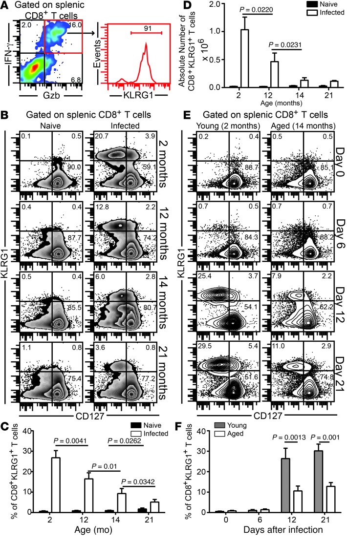
Figure 1. Attrition of effector CD8+KLRG1+ T cells in E. cuniculi–challenged aged mice.
(A) Young mice were orally infected with E. cuniculi spores, and KLRG1 expression was assessed at day 12 after infection in IFN-γ+Gzb+ splenic CD8+ T cells. (B–D) Frequency (B and C) and absolute number (D) of CD8+KLRG1+ T cells in E. cuniculi–challenged animals at 2, 12, 14, and 21 months of age. (E and F) KLRG1+ CD8+ kinetic was assessed in young and 14-month-old mice at days 0, 6, 12, and 21 after infection. Data represent 2 experiments with at least 4 mice per group. Numbers in dot plots and histograms denote percentages.

