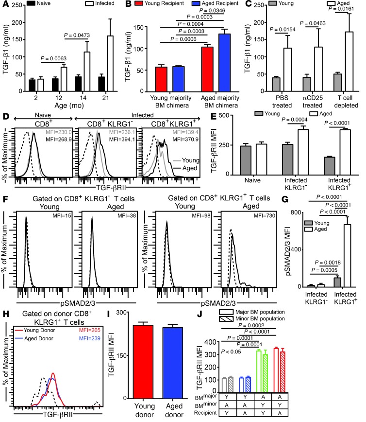
Figure 4. Plasma TGF-β1 is highly elevated in E. cuniculi–challenged aged mice.
(A) TGF-β1 plasma levels in parasite-infected mice of various ages at day 12 after infection. (B) TGF-β plasma levels in E. cuniculi–challenged young majority and aged majority BM chimeras generated in young or aged recipients. (C) Plasma collected from young (6–8 weeks old) or aged (14–15 months old) mice treated with PBS, with anti-CD25, or with anti-thymocyte to deplete T cells was assayed for TGF-β. (D and E) TGF-βRII expression was measured on splenic CD8+ subsets in young (6–8 weeks old) and aged (14–15 months old) mice by polychromatic flow cytometry. (F and G) Direct ex vivo SMAD2/3 phosphorylation on splenic CD8+ subsets. (H and I) Young (CD90.1; 6–8 weeks old) and aged (CD90.2; 14 months old) CD8+ T cells from naive donors were adoptively transferred into young Cd8–/– recipients, followed by parasite challenge. TGF-βRII expression was evaluated in the recipients on donor effector CD8+KLRG1+ T cells in spleen. (J) TGF-βRII expression levels on splenic effector CD8+KLRG1+ T cells in young majority or aged majority BM chimeras formed in young or aged recipients. Y, young; A, aged. Data represent 2 experiments with 4 mice per group. Numbers in histograms denote MFI.

