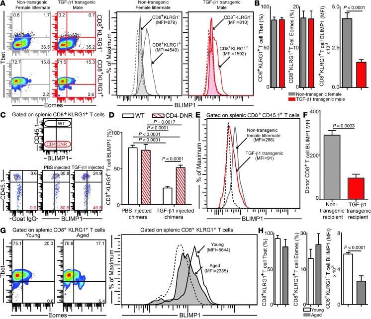
Figure 8. Highly elevated TGF-β levels downregulate BLIMP1 expression on effector CD8+KLRG1+ T cells in the E. cuniculi model.
(A and B) Expression levels of transcription factors Tbet, Eomes, and BLIMP1 in splenic CD8+KLRG1+ and CD8+KLRG1– T cells in young TGF-β1 transgenic mice and littermate controls. (C and D) Acutely infected Young WT:Young CD4-DNR BM chimeras were treated i.v. with 500 ng murine TGF-β1 or PBS. BLIMP1 level was then evaluated in KLRG1+ effectors at day 12 after infection. (E and F) YFP+CD8+ effectors were sorted from E. cuniculi–challenged IFN-γ reporter mice (CD45.1; day 12 after infection) and adoptively transferred to Alb–TGF-β1 recipients or littermate controls at day 7 after infection. BLIMP1 expression in donor cells was assessed 5 days after transfer. (G and H) BLIMP1 level in KLRG1+ CD8+ T cells in young or aged mice at day 12 after infection. Data represent at least 2 experiments with 4–5 mice per group. Numbers in dot plots represent percentages; numbers in histograms represent MFI.

